
ADC programs investigating target-related effects, tissue distribution or tumor-cell killing. Normal-tissue risk assessment can be added where relevant.
R&D service
ADC distribution & efficacy in organoids
Combine target expression, spatial distribution, cytotoxicity and bystander-effect readouts in an organoid study matched to the candidate ADC.
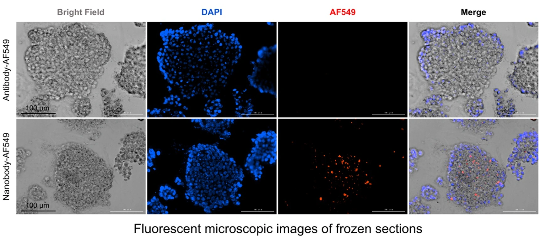 View image
ADC study design
Antibody/nanobody distribution imaging and ADC activity studies
Breast cancer PDO studies combine frozen-section distribution of AF549-labeled antibody/nanobody formats with morphology and viability after ADC treatment. A new candidate program can add target profiling, imaging controls, dedicated penetration quantification and killing endpoints.
Study focus
What this study can help you decide
- 01How are candidate molecules distributed across organoid sections?
- 02How do target expression and cytotoxicity relate?
- 03When are penetration, bystander or immune endpoints valuable?
STUDY APPROACH
Match the model and assays to your program
Select tumor PDO or PDO 2.0 by target and indication, adding normal kidney, lung and GI models or adjacent non-tumor liver organoids to assess organ-specific risk.
Target and histology, spatial distribution, viability and apoptosis; penetration studies can add serial sections, z-stacks and distance/intensity analysis when required.
Unconjugated antibody, free payload, isotype or non-targeting controls and mechanism-appropriate positive controls.
Compare tissue distribution and effect profiles, then prioritize efficacy or safety signals for follow-up.
Study workflow
Relate target expression to ADC distribution, killing and bystander effects in human organoids
ADC studies evaluate target heterogeneity, candidate spatial distribution and subsequent killing together, with penetration quantification, bystander or immune endpoints selected around payload and mechanism.
Target and model assessment
Use histology, IHC or IF to assess target expression and spatial heterogeneity alongside model morphology, viability and the candidate mechanism.
Labeling, spatial imaging and controls
Define labeling, incubation and imaging conditions around molecule properties, then select isotype, unconjugated-antibody, competition or target-negative controls; add serial sections or z-stacks and distance/intensity analysis when penetration quantification is required.
Killing and mechanism endpoints
Relate spatial distribution to dose response, viability, apoptosis and histology, then select bystander or immune endpoints around the study mechanism.
ADC comparison and reporting
Compare candidates under consistent conditions and deliver original images, quantitative methods, analysis and interpretation of target–distribution–effect relationships.
Study images and readouts
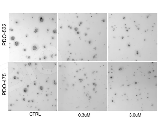
Morphology and dose response after ADC treatment
Bright-field morphology and viability curves show how two breast cancer PDO models respond across ADC concentrations, informing dose selection, model comparison and follow-on studies.
 View image
Post-treatment morphology: bright-field views under control, 0.3 μM and 3.0 μM conditions provide tissue context for viability readouts.
TRiCBIO breast cancer PDO ADC study dataView antibody distribution and ADC response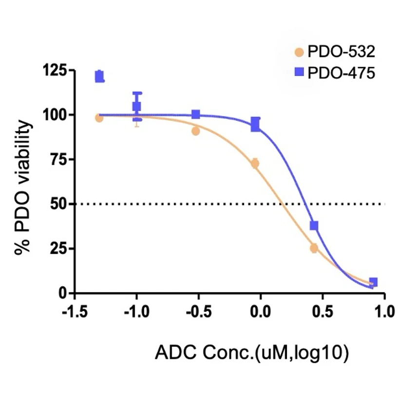 View image
Dose response: two PDO models show different viability profiles across a candidate ADC concentration range, informing replication and follow-on sample studies.
TRiCBIO breast cancer PDO ADC study dataView antibody distribution and ADC responseExpected study outputs
What you receive
Target and spatial distribution
Images of target expression, tissue architecture and candidate spatial distribution, with penetration quantification when specified in the study plan.
Killing and mechanism evidence
An analysis of viability, apoptosis and histology alongside spatial findings, with bystander readouts when included in the study plan.
ADC comparison report
Raw data, analytical methods and a report comparing candidates and explaining the findings.
Relevant model systems
Recommended model systems
Start with target expression and spatial heterogeneity. Tailor labeling, dose, timing, controls, tissue distribution and killing endpoints to the antibody format, linker–payload mechanism, expected exposure and comparison goal. Add bystander or immune readouts around the mechanism.
PROJECT DISCUSSION
Connect target, distribution and killing
Tell us the target, ADC design, intended tumor model and the distribution, killing or bystander question. We’ll configure imaging and efficacy endpoints around the mechanism.