Models, spatial imaging and functional endpoints for ADC studies
Start with target and model selection, then combine section-level spatial distribution, post-treatment morphology and dose response; add dedicated imaging and quantification when the study needs an intact-organoid view of penetration.
Match models and complementary endpoints to the candidate
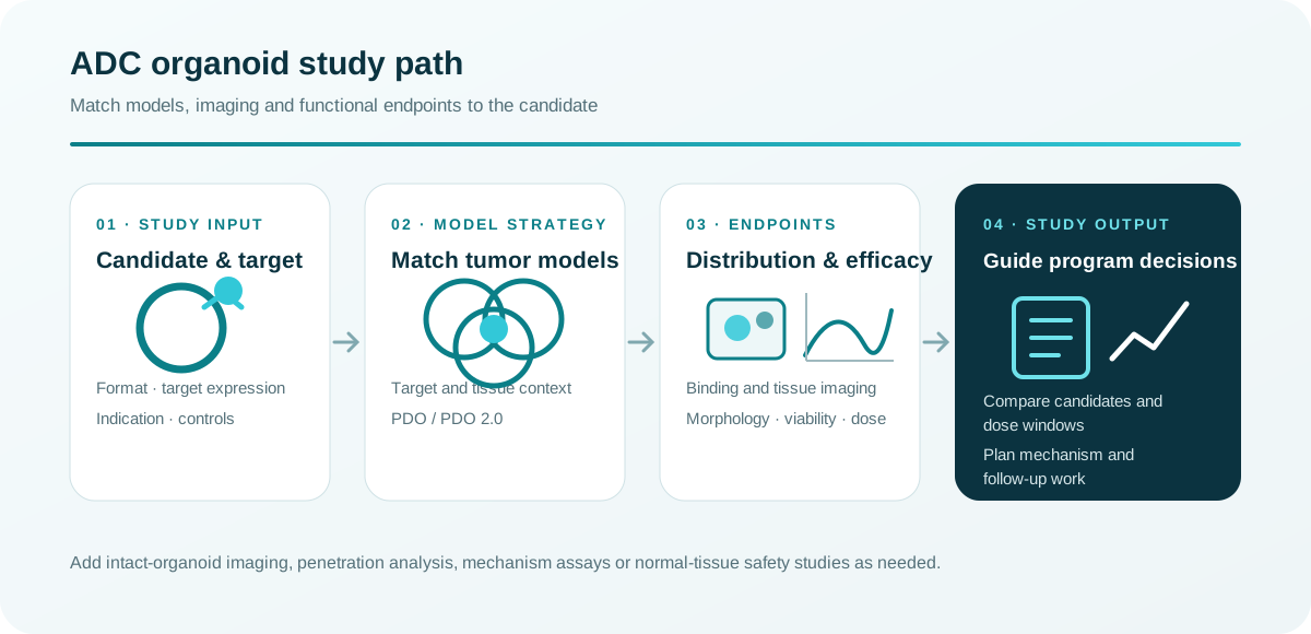
Choose models from candidate format, target and tissue context, then combine spatial distribution, morphology, viability and dose response, with mechanism or normal-tissue safety studies added as needed.
Organizes imaging, efficacy and follow-up validation into one connected study plan
Connect spatial-distribution imaging with efficacy endpoints
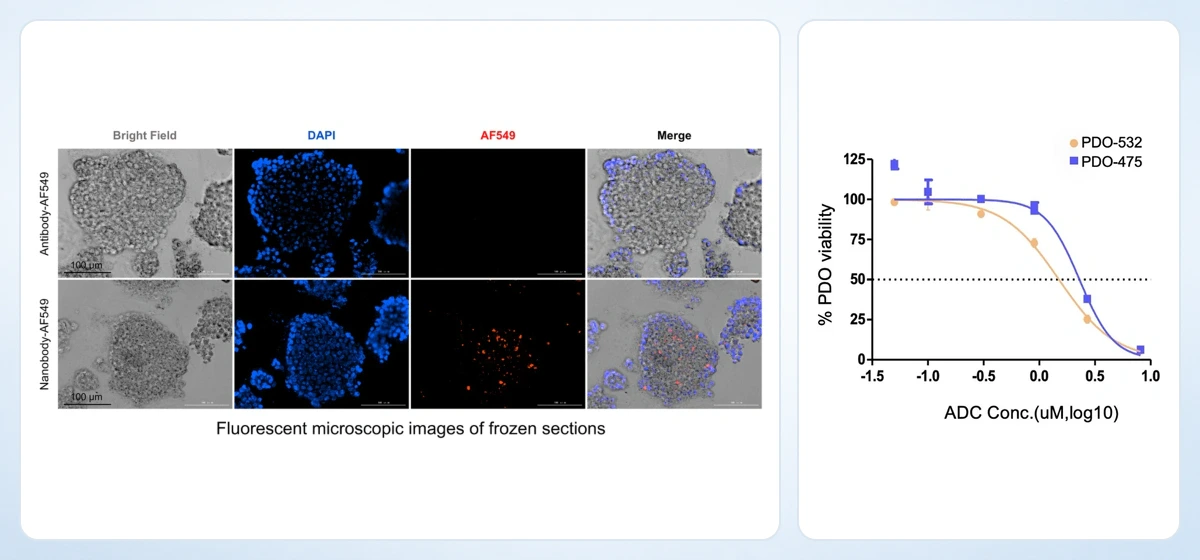
Brings together frozen-section distribution data and complementary PDO morphology and dose-response data to show how imaging and functional endpoints work together in an ADC program.
Guides molecular-format, imaging, dose-window and candidate-comparison planning
01
Define the target and model context
Target expression, tissue architecture and candidate mechanism jointly determine the model and controls.
02
Map relative spatial distribution in PDO frozen sections
Labeling and imaging compare relative spatial distribution in PDO frozen sections against target expression; serial sections or z-stacks and distance/intensity analysis can be added when penetration requires quantification.
03
Interpret cell killing with mechanism-focused endpoints
Design viability, apoptosis, histology and bystander-effect endpoints together to build a coherent response profile.
Raw imaging, quantitative outputs, analysis methods and a study report are delivered with the labels, doses and time points specified in the study plan.
Models, spatial imaging and functional endpoints for ADC studies
Start with target and model selection, then combine section-level spatial distribution, post-treatment morphology and dose response; add dedicated imaging and quantification when the study needs an intact-organoid view of penetration.
Match models and complementary endpoints to the candidate
Choose models from candidate format, target and tissue context, then combine spatial distribution, morphology, viability and dose response, with mechanism or normal-tissue safety studies added as needed.
Organizes imaging, efficacy and follow-up validation into one connected study plan
Connect spatial-distribution imaging with efficacy endpoints
Brings together frozen-section distribution data and complementary PDO morphology and dose-response data to show how imaging and functional endpoints work together in an ADC program.
Guides molecular-format, imaging, dose-window and candidate-comparison planning
01
Define the target and model context
Target expression, tissue architecture and candidate mechanism jointly determine the model and controls.
02
Map relative spatial distribution in PDO frozen sections
Labeling and imaging compare relative spatial distribution in PDO frozen sections against target expression; serial sections or z-stacks and distance/intensity analysis can be added when penetration requires quantification.
03
Interpret cell killing with mechanism-focused endpoints
Design viability, apoptosis, histology and bystander-effect endpoints together to build a coherent response profile.
Raw imaging, quantitative outputs, analysis methods and a study report are delivered with the labels, doses and time points specified in the study plan.