
Tumor sample context
Tumor type, collection site, sample condition, expected timing and available ethics documentation.
Established from primary or metastatic colorectal tumors to compare drug response, tissue features and inter-sample differences.
Primary or metastatic colorectal cancer tissue, with sample condition and modeling strategy integrated into study design
PDO
TRiCBIO colorectal cancer PDO histology and drug-response study data
Model characterization
Source-tissue and organoid imaging plus lineage/identity markers characterize the model; functional readouts are selected for the candidate mechanism and study objective.
CONVENTIONAL PDO BASELINE
Source-tissue H&E, PDO bright-field and PDO H&E establish the morphological and histologic reference for identity, efficacy and mechanism studies.
 View image
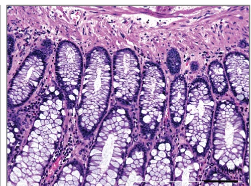
Colorectal source-tissue reference: H&E shows epithelial, glandular and stromal context for PDO morphology and identity assessment.
Model & assay data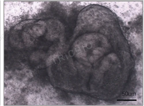 View image
Culture morphology: bright-field imaging documents 3D growth and culture state for subsequent histology and treatment studies.
Model & assay data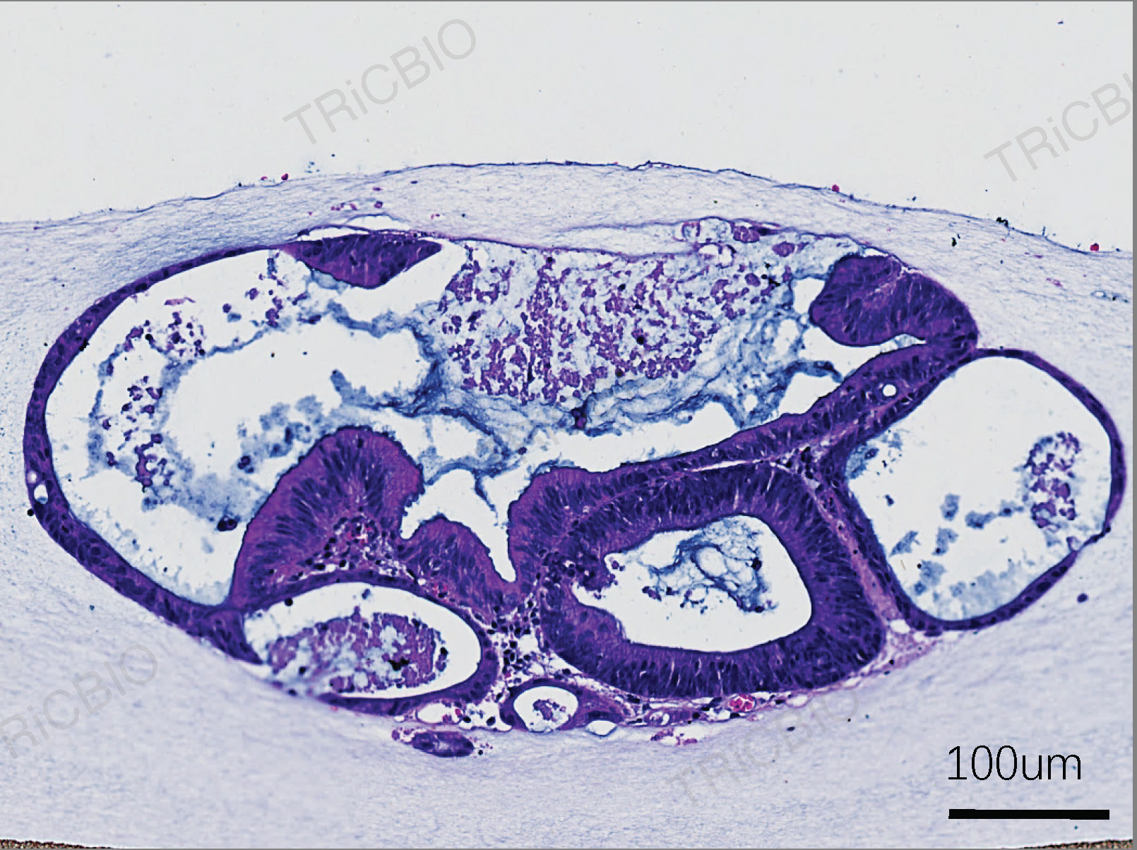 View image
PDO histology: PDO H&E and the source-tissue reference show morphological correspondence and guide identity and functional assays.
Model & assay dataPDO 2.0
Complementary PDO 2.0 data add histology, bright-field imaging and CD3, CD20, Pan-CK and Ki67 profiling when a study requires tissue and immune-context endpoints.
 View image
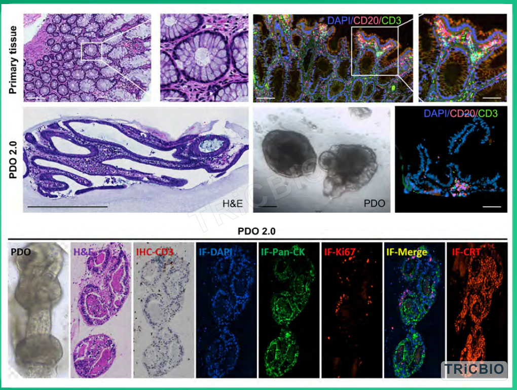
PDO 2.0 characterization: source-tissue and PDO histology, bright-field imaging and CD3, CD20, Pan-CK and Ki67 profiling show epithelial structure and detectable sample-derived immune-cell populations, with the analytical panel configured for each sample.
Study dataStudy planning
Tumor type, sample condition, candidate and intended comparison guide the culture format, controls and assays.
Tumor type, collection site, sample condition, expected timing and available ethics documentation.
Modality, dosing plan, controls and the response or comparison the study should address.
Planned histology, flow, molecular or functional assays, together with preferred data and report formats.
Study design
Primary or metastatic site, prior treatment and sample state can all affect the model. Define whether the study ranks candidates, compares combinations or explores inter-sample response differences.
Review primary or metastatic origin, pathology, prior therapy and sample count, then use consistent analysis criteria to separate source variation from treatment effects.
Use morphology, H&E, epithelial identity and study-relevant immune profiling to select models for a consistent comparison.
Run single-agent or combination studies with consistent dose, timing, controls and analysis, retaining the needed histologic and mechanistic endpoints.
Integrate model QC, response curves and tissue or immune findings to distinguish treatment effects from sample variation, prioritize candidate strategies and define follow-on mechanism studies.
Research applications
Assays & QC
Tissue architecture and epithelial identity establish the baseline for drug-response studies; immune-focused programs can add PDO 2.0 / ALI.
Study & delivery
Choose a supplied model or a TRiCBIO-run efficacy study. Typical deliverables include histology, viability data and a project report.
Related solutions
PROJECT DISCUSSION
Tumor type, sample status, candidate profile and comparison endpoints determine the culture format, controls, assay panel and deliverables for the colorectal cancer PDO.
Established from primary or metastatic colorectal tumors to compare drug response, tissue features and inter-sample differences.
Primary or metastatic colorectal cancer tissue, with sample condition and modeling strategy integrated into study design
PDO
TRiCBIO colorectal cancer PDO histology and drug-response study data
Model characterization
Source-tissue and organoid imaging plus lineage/identity markers characterize the model; functional readouts are selected for the candidate mechanism and study objective.
CONVENTIONAL PDO BASELINE
Source-tissue H&E, PDO bright-field and PDO H&E establish the morphological and histologic reference for identity, efficacy and mechanism studies.
View imageColorectal source-tissue reference: H&E shows epithelial, glandular and stromal context for PDO morphology and identity assessment.
Model & assay dataView imageCulture morphology: bright-field imaging documents 3D growth and culture state for subsequent histology and treatment studies.
Model & assay dataView imagePDO histology: PDO H&E and the source-tissue reference show morphological correspondence and guide identity and functional assays.
Model & assay dataPDO 2.0
Complementary PDO 2.0 data add histology, bright-field imaging and CD3, CD20, Pan-CK and Ki67 profiling when a study requires tissue and immune-context endpoints.
View imagePDO 2.0 characterization: source-tissue and PDO histology, bright-field imaging and CD3, CD20, Pan-CK and Ki67 profiling show epithelial structure and detectable sample-derived immune-cell populations, with the analytical panel configured for each sample.
Study dataStudy planning
Tumor type, sample condition, candidate and intended comparison guide the culture format, controls and assays.
Tumor type, collection site, sample condition, expected timing and available ethics documentation.
Modality, dosing plan, controls and the response or comparison the study should address.
Planned histology, flow, molecular or functional assays, together with preferred data and report formats.
Study design
Primary or metastatic site, prior treatment and sample state can all affect the model. Define whether the study ranks candidates, compares combinations or explores inter-sample response differences.
Review primary or metastatic origin, pathology, prior therapy and sample count, then use consistent analysis criteria to separate source variation from treatment effects.
Use morphology, H&E, epithelial identity and study-relevant immune profiling to select models for a consistent comparison.
Run single-agent or combination studies with consistent dose, timing, controls and analysis, retaining the needed histologic and mechanistic endpoints.
Integrate model QC, response curves and tissue or immune findings to distinguish treatment effects from sample variation, prioritize candidate strategies and define follow-on mechanism studies.
Research applications
Assays & QC
Tissue architecture and epithelial identity establish the baseline for drug-response studies; immune-focused programs can add PDO 2.0 / ALI.
Study & delivery
Choose a supplied model or a TRiCBIO-run efficacy study. Typical deliverables include histology, viability data and a project report.
Related solutions
PROJECT DISCUSSION
Tumor type, sample status, candidate profile and comparison endpoints determine the culture format, controls, assay panel and deliverables for the colorectal cancer PDO.