
Consider PDO 1.0
Confirm model identity, epithelial phenotype, target and drug-activity endpoints.
Compare the biology each culture system can preserve and measure, then select or combine them around mechanism, endpoints and sample context.
CHOOSING THE RIGHT MODEL
Choose the model around the candidate mechanism, the tissue and cell context that matter, and the evidence needed for the next study step.
HOW TO COMBINE
MODEL CONTEXT
Culture morphology and whole-mount immunofluorescence show the state of an epithelial PDO, while tissue-to-PDO histology and immunohistochemistry add structural and lineage context. Select or combine these views around the candidate mechanism, sample and study endpoints.
 View image
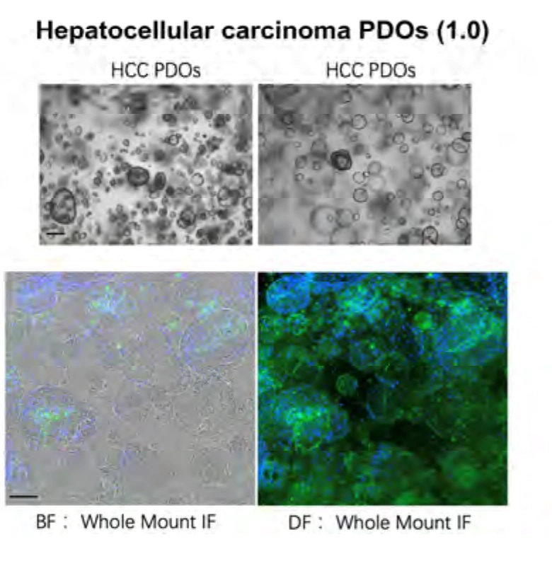
Conventional PDO culture: bright-field and whole-mount immunofluorescence show model morphology and cell state, supporting characterization and downstream endpoint design.
Study data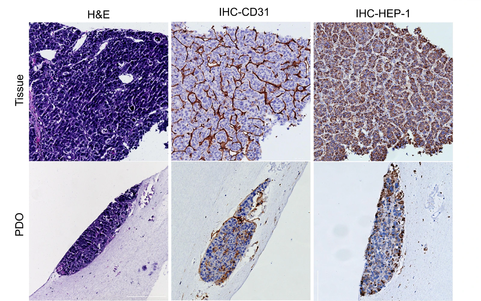 View image
Tissue and model profiling: paired H&E, CD31 and HEP-1 immunohistochemistry in source tissue and PDO shows tissue architecture, vascular features and hepatic lineage markers, supporting model identity and the selection of target and mechanism readouts.
Model & assay dataRelated models and services
PROJECT DISCUSSION
Share your project goal and available materials. Our scientists will recommend models, controls, assays and a practical first study.
Compare the biology each culture system can preserve and measure, then select or combine them around mechanism, endpoints and sample context.
CHOOSING THE RIGHT MODEL
Choose the model around the candidate mechanism, the tissue and cell context that matter, and the evidence needed for the next study step.
HOW TO COMBINE
MODEL CONTEXT
Culture morphology and whole-mount immunofluorescence show the state of an epithelial PDO, while tissue-to-PDO histology and immunohistochemistry add structural and lineage context. Select or combine these views around the candidate mechanism, sample and study endpoints.
View imageConventional PDO culture: bright-field and whole-mount immunofluorescence show model morphology and cell state, supporting characterization and downstream endpoint design.
Study dataView imageTissue and model profiling: paired H&E, CD31 and HEP-1 immunohistochemistry in source tissue and PDO shows tissue architecture, vascular features and hepatic lineage markers, supporting model identity and the selection of target and mechanism readouts.
Model & assay dataRelated models and services
PROJECT DISCUSSION
Share your project goal and available materials. Our scientists will recommend models, controls, assays and a practical first study.