01Source tissue
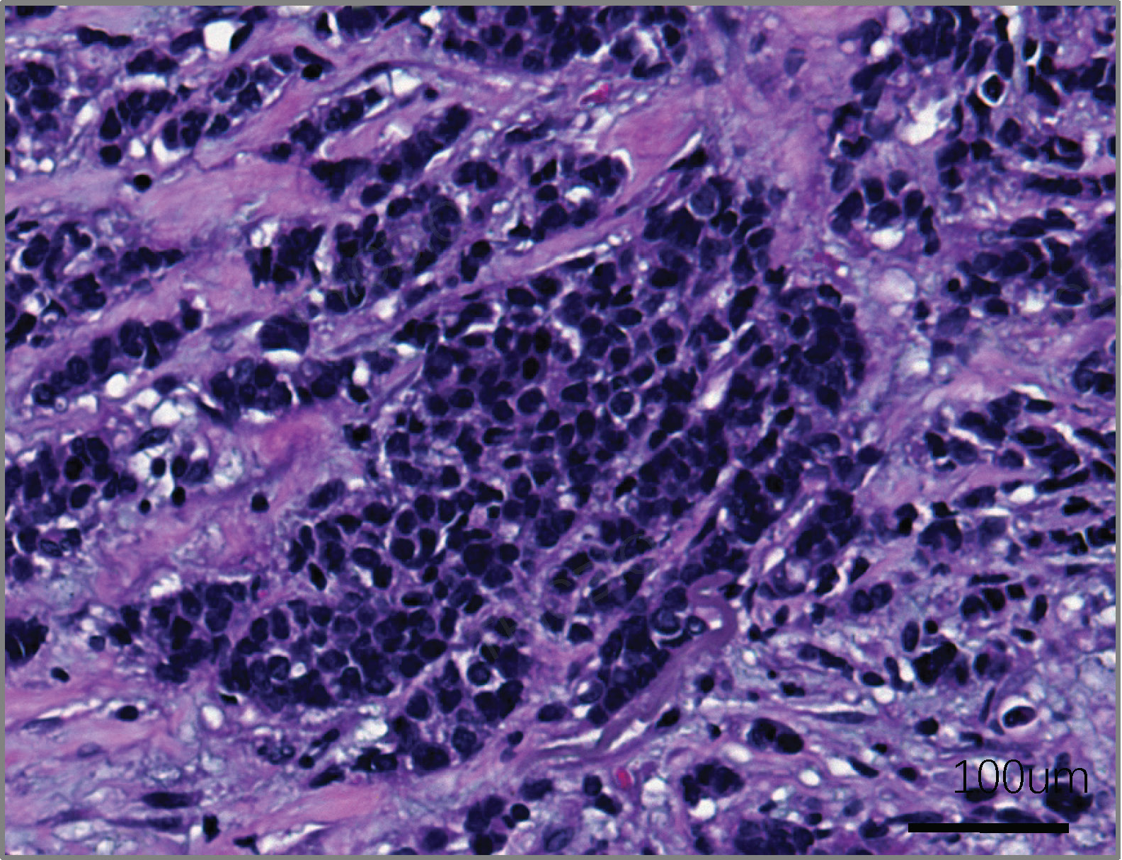
Source-tissue H&E morphology
Source-tissue H&E provides the histological reference for subsequent PDO morphology and histology.
Establishes a histologic reference for PDO quality assessmentAssess ER and Ki67 phenotypes alongside source-tissue/PDO histology to establish model identity and select efficacy endpoints.
SUBTYPE & TISSUE IDENTITY
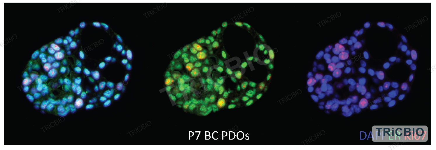
ER and Ki67 show receptor-associated and proliferative phenotypes; source-tissue and PDO morphology and histology then guide model and endpoint selection for subtype-focused efficacy studies.
 View image
Receptor and proliferation phenotype: DAPI, ER and Ki67 show nuclear, receptor-associated and proliferative features in this breast PDO and provide a baseline for subtype-informed study design.
Model & assay dataSTUDY EVIDENCE
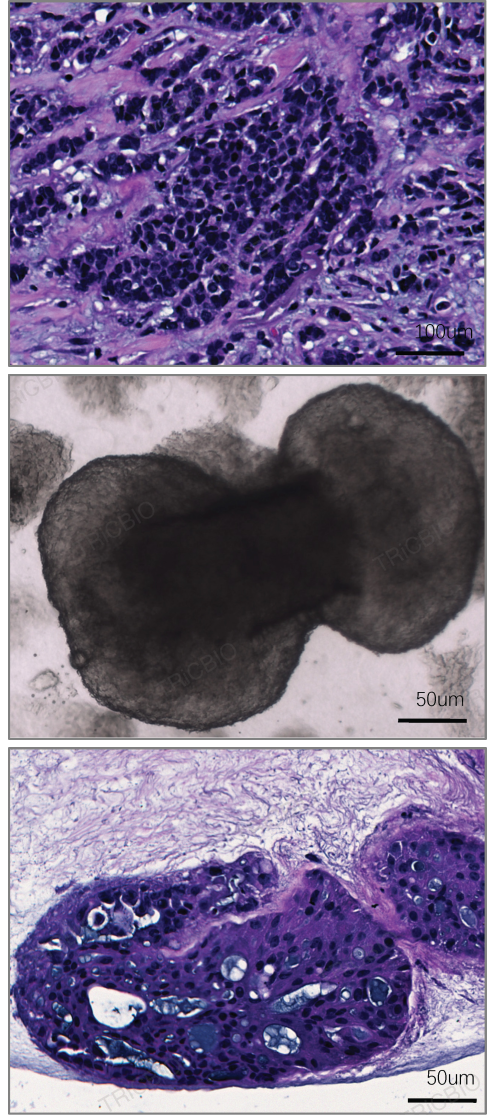
Source-tissue H&E, PDO bright-field imaging and PDO histology establish morphological correspondence and inform receptor, proliferation and target profiling.
Source-tissue H&E provides the histological reference for subsequent PDO morphology and histology.
Establishes a histologic reference for PDO quality assessment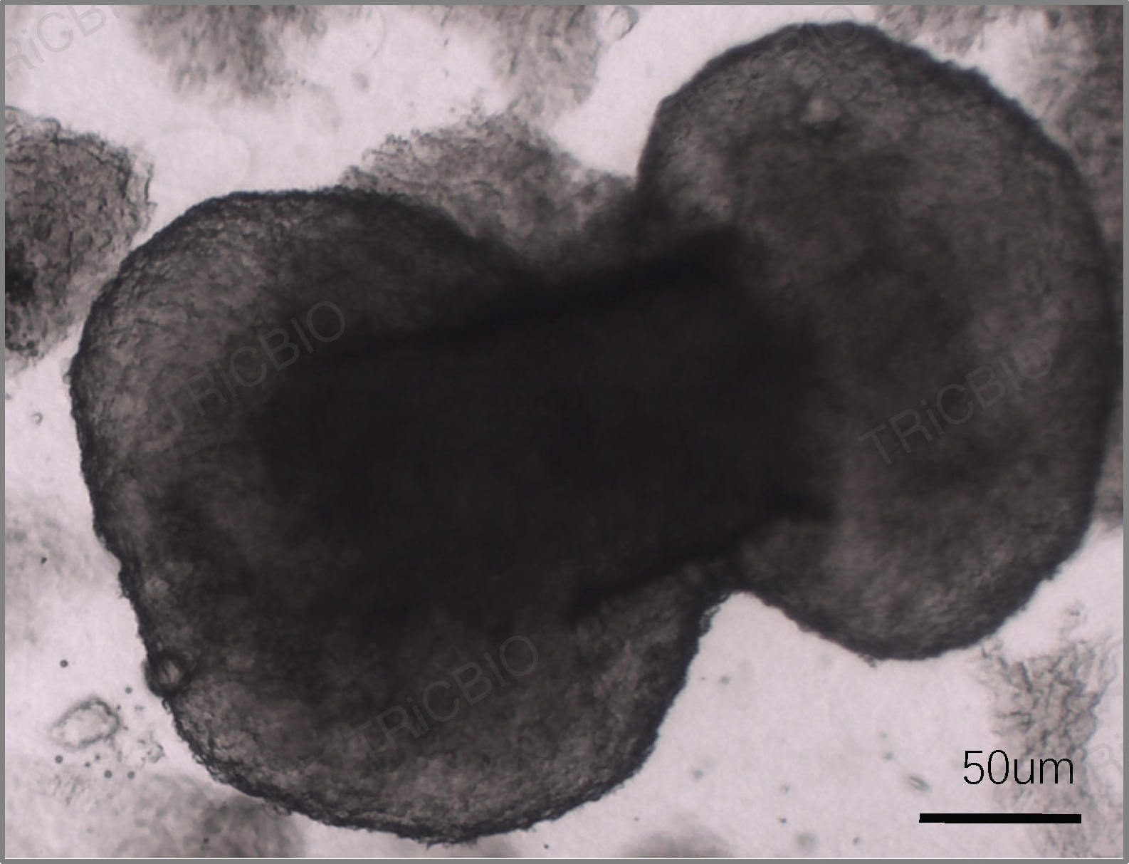
Bright-field imaging documents 3D growth morphology before histology and downstream treatment studies.
Shows culture status before further profiling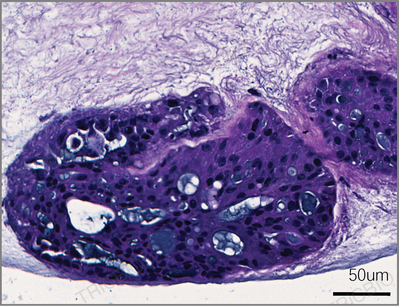
PDO H&E is interpreted with the source-tissue reference to assess morphology; molecular concordance is evaluated with markers and omics assays selected for the study.
Informs receptor, proliferation and target assaysINTERPRETATION
Source-tissue and PDO morphological comparisons plus receptor profiling clarify sample context and model subtype phenotype.
Morphological correspondence, receptor phenotype and culture state support model selection, with long-term stability confirmed through passage and batch QC.
Use subtype and target context to select the most relevant efficacy, ADC, combination or mechanism study.
Related models and services
Study planning
Model characterization, subtype-focused studies or candidate evaluation
Pathology subtype, sample context, target background and downstream objective
Culture and tissue comparison, subtype-associated profiling and model selection for the intended study
Bright-field and histology images, marker results, QC assessment and recommended study conditions
Define the subtype- or target-informed study
Receptor status, culture stability, candidate mechanism and inter-sample variation
PROJECT DISCUSSION
Tell us which model, assay or finding you want to extend. Our scientists will adapt the approach to your candidate, samples and decision point.
Assess ER and Ki67 phenotypes alongside source-tissue/PDO histology to establish model identity and select efficacy endpoints.
SUBTYPE & TISSUE IDENTITY
ER and Ki67 show receptor-associated and proliferative phenotypes; source-tissue and PDO morphology and histology then guide model and endpoint selection for subtype-focused efficacy studies.
View imageReceptor and proliferation phenotype: DAPI, ER and Ki67 show nuclear, receptor-associated and proliferative features in this breast PDO and provide a baseline for subtype-informed study design.
Model & assay dataSTUDY EVIDENCE
Source-tissue H&E, PDO bright-field imaging and PDO histology establish morphological correspondence and inform receptor, proliferation and target profiling.
Source-tissue H&E provides the histological reference for subsequent PDO morphology and histology.
Establishes a histologic reference for PDO quality assessmentBright-field imaging documents 3D growth morphology before histology and downstream treatment studies.
Shows culture status before further profilingPDO H&E is interpreted with the source-tissue reference to assess morphology; molecular concordance is evaluated with markers and omics assays selected for the study.
Informs receptor, proliferation and target assaysINTERPRETATION
Source-tissue and PDO morphological comparisons plus receptor profiling clarify sample context and model subtype phenotype.
Morphological correspondence, receptor phenotype and culture state support model selection, with long-term stability confirmed through passage and batch QC.
Use subtype and target context to select the most relevant efficacy, ADC, combination or mechanism study.
Related models and services
Study planning
Model characterization, subtype-focused studies or candidate evaluation
Pathology subtype, sample context, target background and downstream objective
Culture and tissue comparison, subtype-associated profiling and model selection for the intended study
Bright-field and histology images, marker results, QC assessment and recommended study conditions
Define the subtype- or target-informed study
Receptor status, culture stability, candidate mechanism and inter-sample variation
PROJECT DISCUSSION
Tell us which model, assay or finding you want to extend. Our scientists will adapt the approach to your candidate, samples and decision point.