
Target and model assessment
Assess target location, intensity and heterogeneity, then define model-selection criteria.
Application
Evaluate target expression, effector-cell conditions, dynamic killing and immune activation together in human 3D tumor models.
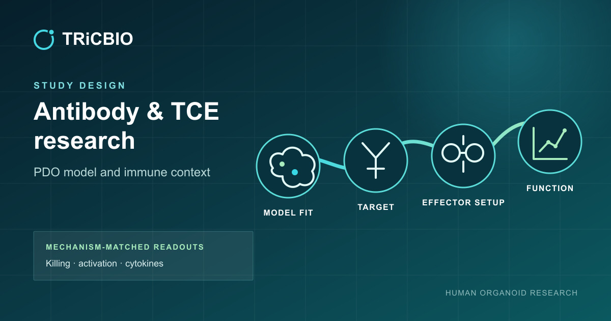
APPLICATION PATH
Conventional PDOs support standardized tumor-killing comparisons. When tissue architecture or sample-associated immune context matters, PDO 2.0 / ALI can add that layer alongside mechanism-matched effector cells and functional endpoints.
PDAC PDO 2.0 model and immune context
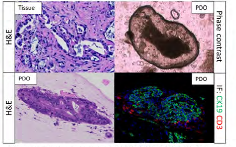
PDAC source-tissue/PDO histology, bright-field imaging and CK19/CD3 immunofluorescence establish epithelial and T-cell context; renal PDO 2.0 evidence adds PanCK/CD3 signals across culture time. Candidate studies can then add effector-cell, activation, killing and cytokine assays around the mechanism.
 View image
PDAC model identity and tissue context: source-tissue H&E, PDO bright-field and PDO H&E are combined with CK19 and CD3 immunofluorescence to add epithelial and sample-associated T-cell context; measurable components and the assay panel are confirmed during model setup.
TRiCBIO PDAC PDO 2.0 study data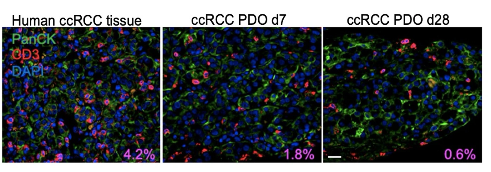 View image
Tissue and model context: PanCK, CD3 and DAPI imaging compares tumor-epithelial and CD3-positive immune-cell signals across the source tissue and PDO culture time points.
TRiCBIO ccRCC PDO 2.0 immune-context study dataContinue to the PDAC and renal PDO 2.0 studies for model context and immune-assay evidence.
Study focus
Study workflow
Assess target location, intensity and heterogeneity, then define model-selection criteria.
Define effector source, E:T ratio, preconditioning and target-relevant positive and negative controls.
Combine live or endpoint imaging, viability/apoptosis, activation markers such as CD107a and cytokines.
Compare potency, response window and activation profile under consistent conditions to build a clear basis for candidate ranking.
Model options
We tailor the effector-cell source, E:T ratio and controls to the target and molecular format so killing, activation and cytokine readouts can support candidate comparison together.
PROJECT DISCUSSION
With the target, molecular format, tumor model and effector-cell conditions defined, we can design the relevant killing and activation comparisons.
Application
Evaluate target expression, effector-cell conditions, dynamic killing and immune activation together in human 3D tumor models.
APPLICATION PATH
Conventional PDOs support standardized tumor-killing comparisons. When tissue architecture or sample-associated immune context matters, PDO 2.0 / ALI can add that layer alongside mechanism-matched effector cells and functional endpoints.
PDAC PDO 2.0 model and immune context
PDAC source-tissue/PDO histology, bright-field imaging and CK19/CD3 immunofluorescence establish epithelial and T-cell context; renal PDO 2.0 evidence adds PanCK/CD3 signals across culture time. Candidate studies can then add effector-cell, activation, killing and cytokine assays around the mechanism.
View imagePDAC model identity and tissue context: source-tissue H&E, PDO bright-field and PDO H&E are combined with CK19 and CD3 immunofluorescence to add epithelial and sample-associated T-cell context; measurable components and the assay panel are confirmed during model setup.
TRiCBIO PDAC PDO 2.0 study dataView imageTissue and model context: PanCK, CD3 and DAPI imaging compares tumor-epithelial and CD3-positive immune-cell signals across the source tissue and PDO culture time points.
TRiCBIO ccRCC PDO 2.0 immune-context study dataContinue to the PDAC and renal PDO 2.0 studies for model context and immune-assay evidence.
Study focus
Study workflow
Assess target location, intensity and heterogeneity, then define model-selection criteria.
Define effector source, E:T ratio, preconditioning and target-relevant positive and negative controls.
Combine live or endpoint imaging, viability/apoptosis, activation markers such as CD107a and cytokines.
Compare potency, response window and activation profile under consistent conditions to build a clear basis for candidate ranking.
Model options
We tailor the effector-cell source, E:T ratio and controls to the target and molecular format so killing, activation and cytokine readouts can support candidate comparison together.
PROJECT DISCUSSION
With the target, molecular format, tumor model and effector-cell conditions defined, we can design the relevant killing and activation comparisons.