01Source tissue
Source-tissue H&E reference
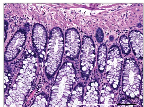
Colorectal source-tissue H&E shows epithelial, glandular and stromal context for PDO QC.
Establishes a histologic reference for model quality assessmentSource-tissue and PDO histology are compared with CD3, CD20, Pan-CK and Ki67 readouts to establish tissue and immune context for the study.
STUDY EVIDENCE
Conventional PDO images establish source-tissue, culture and histological context. PDO 2.0 studies add CD3, CD20, Pan-CK and Ki67 profiling to examine tissue and immune features.
Colorectal source-tissue H&E shows epithelial, glandular and stromal context for PDO QC.
Establishes a histologic reference for model quality assessment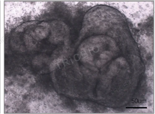
Bright-field imaging documents morphology and culture state; establishment and expansion performance are evaluated through batch QC and culture records.
Shows culture status before histology and treatment studies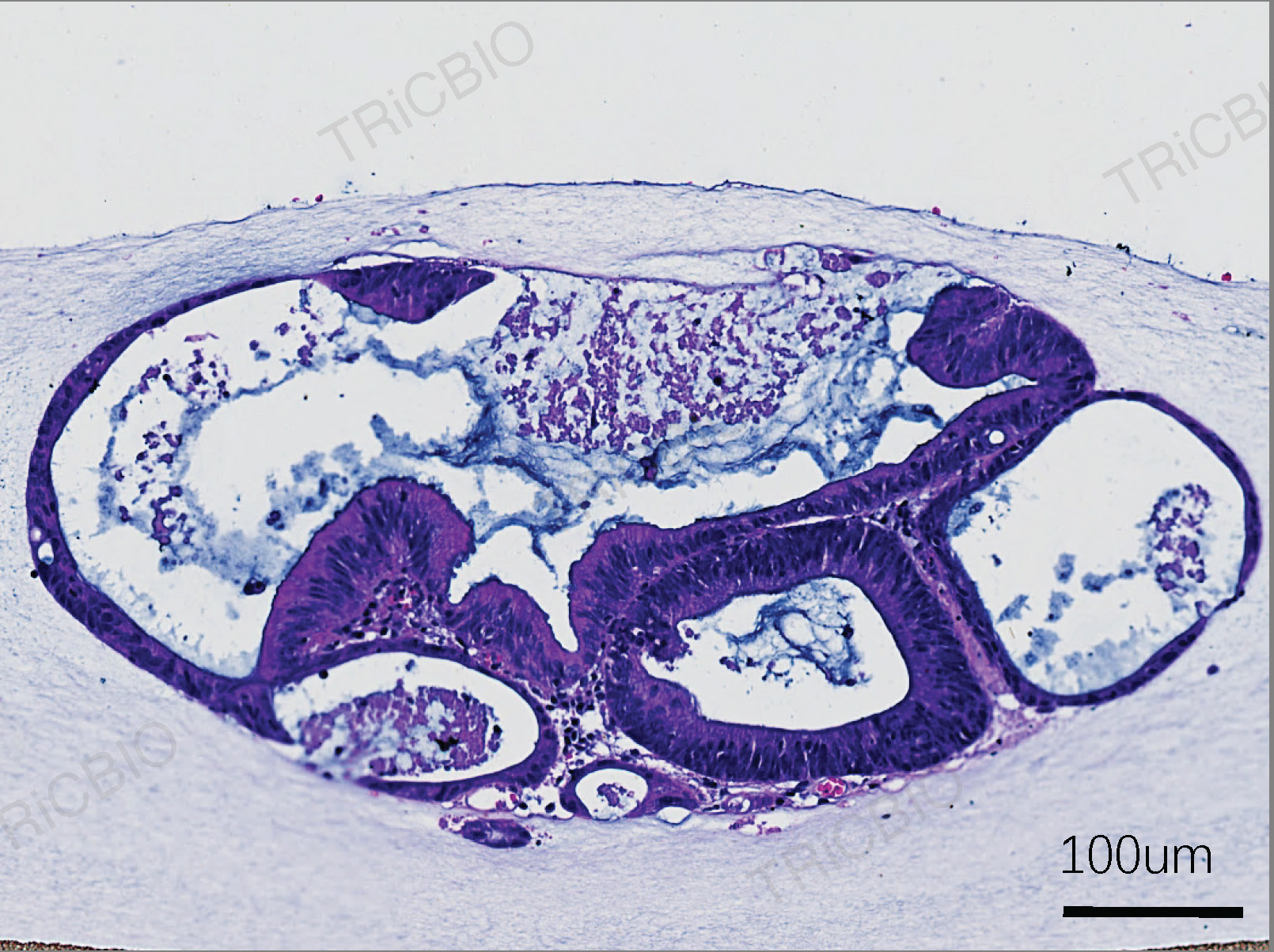
PDO H&E and source tissue show morphological correspondence and combine with pathology and molecular data for tumor-identity assessment.
Informs the combination of identity and functional assays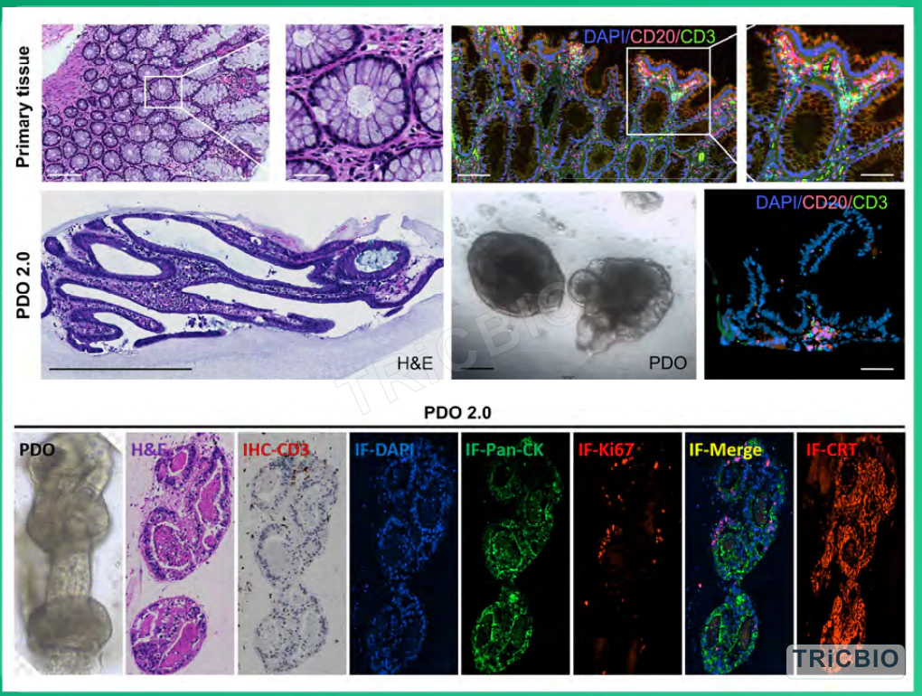
The upper panel compares source tissue and PDO 2.0 through H&E and DAPI/CD20/CD3 imaging, alongside PDO bright-field morphology. The lower panel adds PDO 2.0 bright-field, H&E, IHC-CD3 and IF channels including Pan-CK and Ki67. Tissue architecture, epithelial identity and immune signals inform endpoint selection.
Connects immune-treatment design with tissue and cellular endpointsTogether, culture, histology and immune profiling establish colorectal PDO and PDO 2.0 model QC and guide study design.
INTERPRETATION
Tissue morphology, epithelial identity and measurable immune components establish the model baseline.
New studies use the sample's baseline immune profile to select treatment conditions, assay combinations and observation timing, with serial sampling and replication used to examine subsequent change.
Use the model in efficacy, immune-function, combination or biomarker studies selected for the candidate mechanism.
Related models and services
Study planning
Model characterization, mechanism studies or immuno-oncology planning
Sample pathology, study objective, candidate modality and intended comparisons
Tissue-to-model comparison, baseline characterization and immune assays selected for the study
Study images, assay results, integrated analysis and study-design notes
Define the downstream efficacy or immune-focused study
Immune composition, culture state, candidate mechanism and endpoints for each new sample
PROJECT DISCUSSION
Tell us which model, assay or finding you want to extend. Our scientists will adapt the approach to your candidate, samples and decision point.
Source-tissue and PDO histology are compared with CD3, CD20, Pan-CK and Ki67 readouts to establish tissue and immune context for the study.
STUDY EVIDENCE
Conventional PDO images establish source-tissue, culture and histological context. PDO 2.0 studies add CD3, CD20, Pan-CK and Ki67 profiling to examine tissue and immune features.
Colorectal source-tissue H&E shows epithelial, glandular and stromal context for PDO QC.
Establishes a histologic reference for model quality assessmentBright-field imaging documents morphology and culture state; establishment and expansion performance are evaluated through batch QC and culture records.
Shows culture status before histology and treatment studiesPDO H&E and source tissue show morphological correspondence and combine with pathology and molecular data for tumor-identity assessment.
Informs the combination of identity and functional assaysThe upper panel compares source tissue and PDO 2.0 through H&E and DAPI/CD20/CD3 imaging, alongside PDO bright-field morphology. The lower panel adds PDO 2.0 bright-field, H&E, IHC-CD3 and IF channels including Pan-CK and Ki67. Tissue architecture, epithelial identity and immune signals inform endpoint selection.
Connects immune-treatment design with tissue and cellular endpointsTogether, culture, histology and immune profiling establish colorectal PDO and PDO 2.0 model QC and guide study design.
INTERPRETATION
Tissue morphology, epithelial identity and measurable immune components establish the model baseline.
New studies use the sample's baseline immune profile to select treatment conditions, assay combinations and observation timing, with serial sampling and replication used to examine subsequent change.
Use the model in efficacy, immune-function, combination or biomarker studies selected for the candidate mechanism.
Related models and services
Study planning
Model characterization, mechanism studies or immuno-oncology planning
Sample pathology, study objective, candidate modality and intended comparisons
Tissue-to-model comparison, baseline characterization and immune assays selected for the study
Study images, assay results, integrated analysis and study-design notes
Define the downstream efficacy or immune-focused study
Immune composition, culture state, candidate mechanism and endpoints for each new sample
PROJECT DISCUSSION
Tell us which model, assay or finding you want to extend. Our scientists will adapt the approach to your candidate, samples and decision point.