
Assess the model through tissue morphology, epithelial identity and detectable sample-derived immune-cell populations.
Tumor PDO 2.0
An ALI-based tumor model for studying treatment response alongside source-tissue architecture and immune cells retained from the sample.
CD3CD4CD8CD56CD68
Model source
Patient tumor tissue; model design is tailored to sample condition and study objectives
Culture / format
ALI · 96-well adaptable
Supporting evidence
Published method: the 2018 Cell study of ALI tumor organoids. TRiCBIO evidence: tissue and immune readouts from colorectal and renal PDO 2.0 studies.
Model characterization
Tissue and immune characterization in PDO 2.0
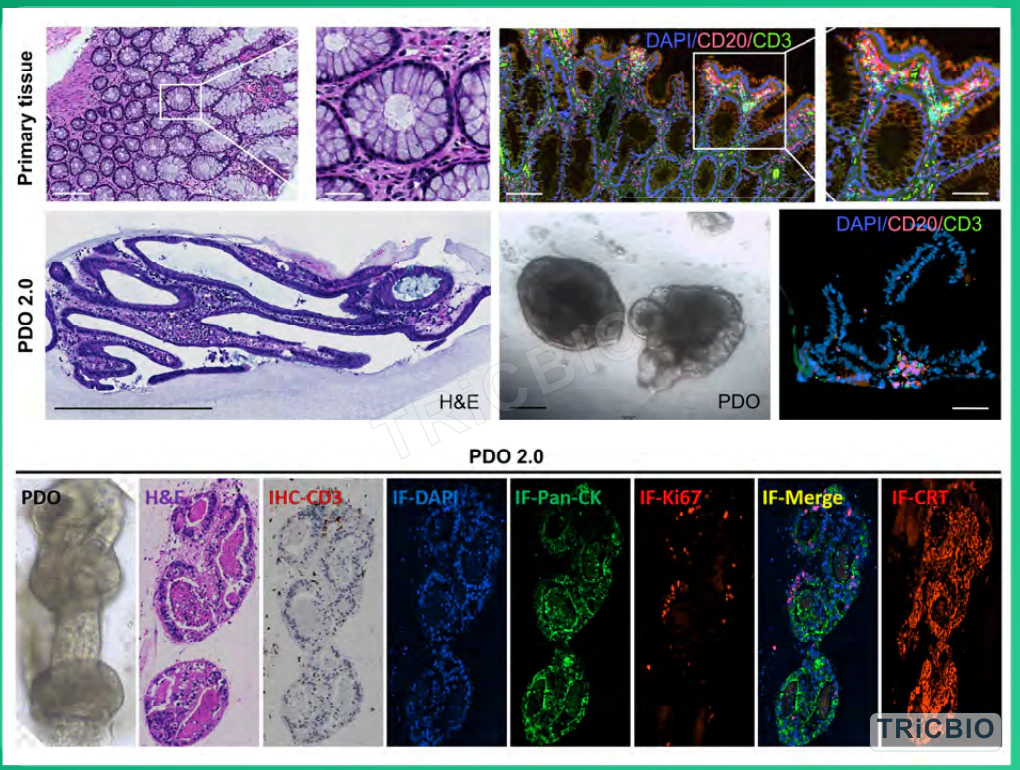
Colorectal and renal PDO 2.0 studies combine histology, bright-field imaging and immune readouts to establish model context and select tumor- and sample-specific endpoints.
 View image
PDO 2.0 characterization: source-tissue and PDO histology, bright-field imaging and CD3, CD20, Pan-CK and Ki67 profiling show epithelial structure and detectable sample-derived immune-cell populations, with the analytical panel configured for each sample.
Study data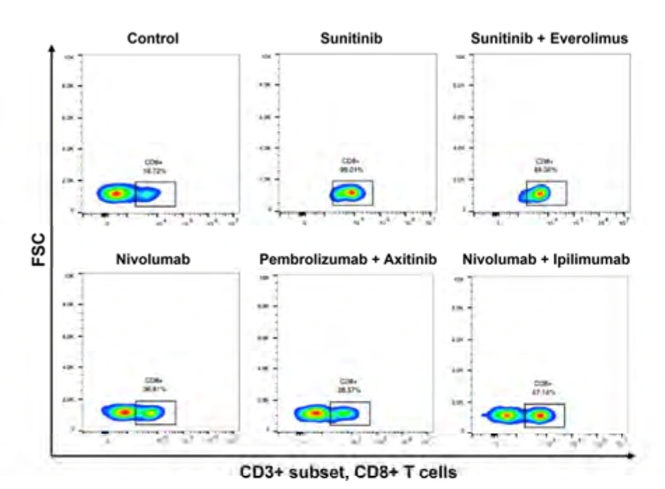
Immune readouts: CD3/CD8 flow cytometry is interpreted with apoptosis measurements to compare cell-death and immune changes across the same treatment conditions.
TRiCBIO RCC PDO 2.0 study dataModel documentation
Model profile and available documentation
Histology, flow, cytokines, treatment response and study-specific immune endpoints.
Use sample immune profiling and culture status to define the study window and select histology, flow and functional assays.
Model and assay summary, model/batch QC, original images or data and study report.
Study design
Connect tissue architecture, immune context and treatment response in PDO 2.0
PDO 2.0 uses ALI culture to study treatment response alongside tissue architecture and the sample’s immune-cell context, supporting immune-mechanism and candidate-comparison studies.
Sample and study design
Bring together tumor type, collection method, tissue condition, timing, ethics documentation and candidate modality to design the ALI study.
Model establishment and baseline QC
Use bright-field imaging, H&E, epithelial identity and immune profiling to confirm the model baseline and study window.
Treatment and multimodal readouts
Select viability, histology, flow, cytokine or TCR readouts around the mechanism and integrate them to interpret complex immune response.
Integrate findings and define follow-up priorities
Integrate model QC, treatment response and immune findings to identify the most relevant observations and priorities for follow-up.
Research applications
Recommended applications
- Checkpoint inhibitors and combinations
- Tumor immune microenvironment profiling
- Efficacy, mechanism and biomarker studies
Assays & QC
Tissue architecture, immune context and treatment readouts
Key markers
CD3CD4CD8CD56CD68
Available assays
- H&E / IHC / IF
- Flow cytometry
- Cytokines
- TCR profiling
QC approach
Tissue morphology, epithelial identity, measurable immune cells and functional state establish the model baseline and study window.
Study & delivery
Model characterization, treatment data and reporting
Programs can cover model establishment and treatment, with raw data, analyses and a study report defined in the project scope.
Related solutions
Related study options
Start a conversation
Start with your project goal
Share the project goal, candidate type and sample context to begin planning the model, assays and first study with our scientific team.
Tumor sample context
Tumor type, collection site, sample condition, expected timing and available ethics documentation.
Candidate and study objective
Modality, dosing plan, controls and the response or comparison the study should address.
Assays and deliverables
Planned histology, flow, molecular or functional assays, together with preferred data and report formats.
PROJECT DISCUSSION
Discuss a PDO 2.0 study
Tumor type, sample status, candidate profile and comparison endpoints determine the culture format, controls, assay panel and deliverables for the PDO 2.0.