
Use histology, epithelial, stromal and immune profiling to select the most relevant efficacy, immune and mechanism endpoints for the PDAC model.
Study resultsCase study
PDAC PDO 2.0: epithelial identity & immune readouts
Compare source-tissue and PDO histology with CK19/CD3 immunofluorescence and CD3/CD8/CD107a flow to assess epithelial identity and immune readouts.
01Study objective02Model & design03Assays & findings04Decision & next step
Model & samplePancreatic ductal adenocarcinoma PDO 2.0 study model
ObjectiveEstablish tissue identity and microenvironment features for downstream efficacy, immune and mechanism studies
Study conditionsBaseline model assessment and candidate conditions selected for the study
AssaysBright-field, histology, epithelial and program-relevant immune markers
Study evidenceTRiCBIO study data and assay readouts
DeliverablesReceive images, assay results and an analysis summary; the report documents the study design, sample size and statistical methods
TISSUE & IMMUNE PROFILING
Epithelial identity and immune profiling
Source-tissue and PDO histology, CK19/CD3 imaging, and CD3/CD8/CD107a flow connect epithelial identity, sample-associated T cells and immune changes across study conditions.
 View image
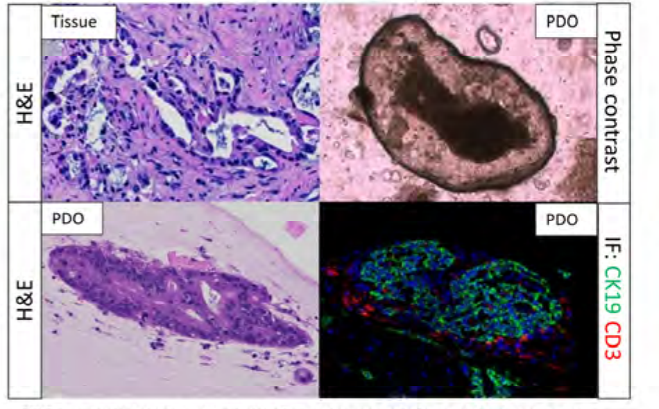
PDAC model identity and tissue context: source-tissue H&E, PDO bright-field and PDO H&E are combined with CK19 and CD3 immunofluorescence to add epithelial and sample-associated T-cell context; measurable components and the assay panel are confirmed during model setup.
TRiCBIO PDAC PDO 2.0 study data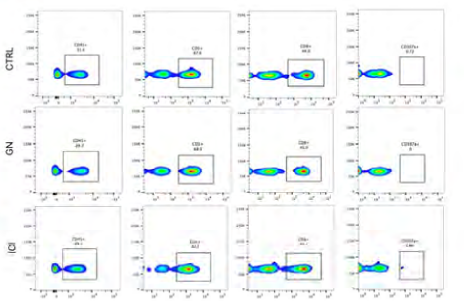 View image
PDAC PDO immune readouts: flow panels assess CD3, CD8 and CD107a populations across conditions, supported by documented treatment definitions, gating, replication and quantitative analysis.
Study dataEstablish PDAC epithelial identity and immune readouts
Source-tissue and PDO morphology establish the model context; CK19, CD3 and flow cytometry then provide complementary views of ductal epithelial identity, sample-associated T cells and treatment-related immune change.
Profile ductal epithelial identity with CK19
Source-tissue H&E, PDO bright-field and PDO H&E assess tissue correspondence. The green immunofluorescence channel is CK19 and adds ductal epithelial context; red CD3 identifies sample-associated T cells.
- Source-tissue and PDO H&E
- PDO bright-field morphology
- CK19 epithelial profiling
- CD3 T-cell profiling
Relate immune readouts to treatment response
CD3, CD8 and CD107a flow cytometry can characterize T-cell composition and changes in degranulation or activation across treatment conditions, with consistent gating, baseline assessment and replication.
Select assay panels by candidate mechanism
Small molecules, ADCs, antibodies and immunotherapies require different endpoints. Once identity is confirmed, studies may add viability, apoptosis, penetration, bystander or immune assays.
- Model and target context
- Viability, apoptosis and histology
- Penetration and bystander endpoints
- CD3/CD8/CD107a flow
Match immune endpoints to candidate mechanism
Model-identity and immune assays work together in one study design. A new program profiles stromal and immune features, then configures gating, controls and replication to compare treatment-related immune change.
INTERPRETATION
Use epithelial and immune readouts to select study endpoints
Establishment, stromal and immune features are assessed against each PDAC sample baseline and extended with candidate mechanism, controls and independent samples.
Set dose, time, functional endpoints and controls around the candidate mechanism, then compare across independent samples.
Related models and services
Choose the right models and services for the next study
PROJECT DISCUSSION
Discuss a study built around your program
Tell us which model, assay or finding you want to extend. Our scientists will adapt the approach to your candidate, samples and decision point.