
Phenotype and comparison groups
Define the most reliable functional phenotype and create comparison groups by response, model feature, dose or time.
R&D service
Relate tissue, functional and molecular changes to the observed phenotype and identify response-associated biomarker candidates for further validation.
 View image
Mechanism and biomarkers across models
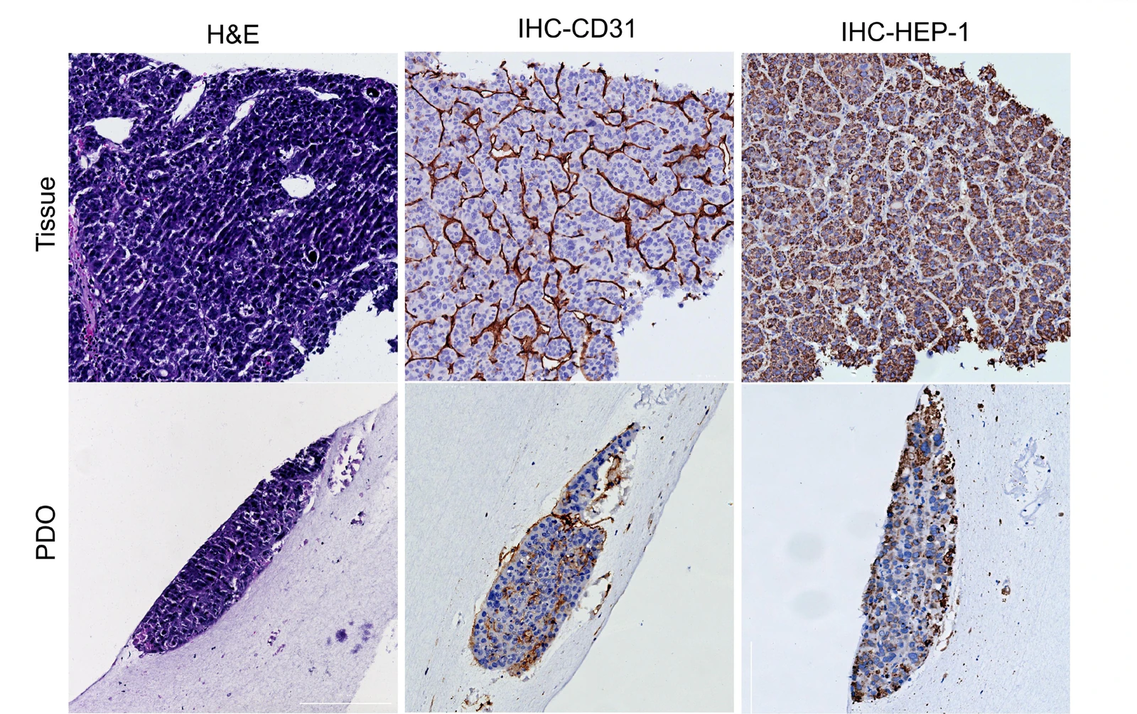
HCC tissue–PDO histology and marker comparisons, kidney-injury qPCR and intestinal tight-junction imaging show how tissue, molecular and functional readouts can be combined around different mechanism questions.
Study focus
Study workflow
Compare baseline features, treatment response and mechanism readouts within the same model and batch context, then confirm key associations in additional samples, with orthogonal methods or repeat perturbation.
Define the most reliable functional phenotype and create comparison groups by response, model feature, dose or time.
Select histology, imaging, flow, qPCR, protein or secreted readouts around the candidate pathway so every readout serves the scientific question.
Compare baseline features, treatment response and mechanism readouts within model and batch context to identify reproducible response-associated candidates.
Confirm key findings in additional samples, with orthogonal methods or repeat perturbation, then assess candidate-marker reproducibility and its relevance to mechanism or response groups.
Study images and readouts
Use tissue images, functional readouts and molecular assays to understand model identity, treatment response and differences across candidate strategies.
 View image
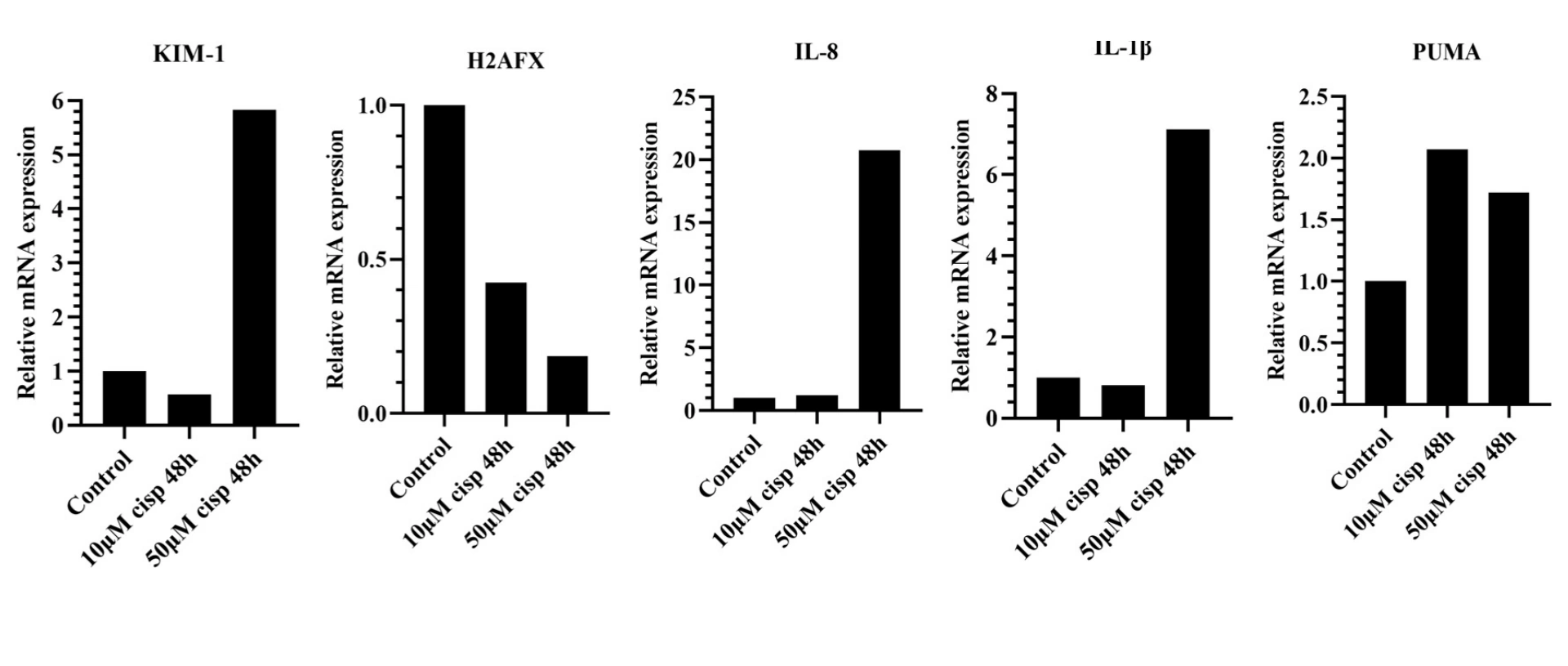
H9-derived | cisplatin molecular response: RT-qPCR profiles KIM-1, H2AFX and inflammation/stress-related genes across cisplatin conditions, helping prioritize kidney-injury biomarkers and define follow-on dose, timing and confirmatory assays.
TRiCBIO H9-derived kidney organoid cisplatin studyView the kidney organoid injury study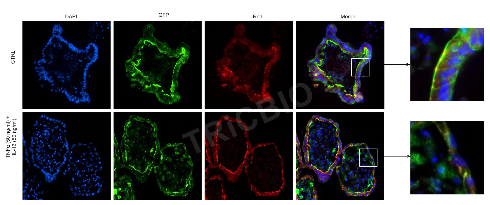 View image
Cytoskeleton and tight junctions: F-actin and ZO-1 immunofluorescence provides epithelial-structure and junction-status readouts.
TRiCBIO mouse intestinal organoid studyView the IBD model and barrier studyExpected study outputs
Functional, morphological, histological and molecular results from the agreed assays, with model and batch information.
Integrate supporting findings, identify associations for follow-up testing and explain how candidate markers were selected.
Prioritized findings, orthogonal confirmation methods and additional sample requirements for the next study.
Relevant model systems
TRiCBIO studies in IBD barrier biology, kidney injury, tumor apoptosis and tumor immunity combine morphological, functional and molecular readouts. Candidate signals can then be advanced through additional samples, orthogonal methods and prespecified analyses to support mechanism and follow-on biomarker research.
PROJECT DISCUSSION
Bring the observed phenotype, comparison groups, available data and mechanism hypothesis. Our scientists will connect molecular, functional and orthogonal assays around that question.
R&D service
Relate tissue, functional and molecular changes to the observed phenotype and identify response-associated biomarker candidates for further validation.
View imageMechanism and biomarkers across models
HCC tissue–PDO histology and marker comparisons, kidney-injury qPCR and intestinal tight-junction imaging show how tissue, molecular and functional readouts can be combined around different mechanism questions.
Study focus
Study workflow
Compare baseline features, treatment response and mechanism readouts within the same model and batch context, then confirm key associations in additional samples, with orthogonal methods or repeat perturbation.
Define the most reliable functional phenotype and create comparison groups by response, model feature, dose or time.
Select histology, imaging, flow, qPCR, protein or secreted readouts around the candidate pathway so every readout serves the scientific question.
Compare baseline features, treatment response and mechanism readouts within model and batch context to identify reproducible response-associated candidates.
Confirm key findings in additional samples, with orthogonal methods or repeat perturbation, then assess candidate-marker reproducibility and its relevance to mechanism or response groups.
Study images and readouts
Use tissue images, functional readouts and molecular assays to understand model identity, treatment response and differences across candidate strategies.
View imageH9-derived | cisplatin molecular response: RT-qPCR profiles KIM-1, H2AFX and inflammation/stress-related genes across cisplatin conditions, helping prioritize kidney-injury biomarkers and define follow-on dose, timing and confirmatory assays.
TRiCBIO H9-derived kidney organoid cisplatin studyView the kidney organoid injury studyView imageCytoskeleton and tight junctions: F-actin and ZO-1 immunofluorescence provides epithelial-structure and junction-status readouts.
TRiCBIO mouse intestinal organoid studyView the IBD model and barrier studyExpected study outputs
Functional, morphological, histological and molecular results from the agreed assays, with model and batch information.
Integrate supporting findings, identify associations for follow-up testing and explain how candidate markers were selected.
Prioritized findings, orthogonal confirmation methods and additional sample requirements for the next study.
Relevant model systems
TRiCBIO studies in IBD barrier biology, kidney injury, tumor apoptosis and tumor immunity combine morphological, functional and molecular readouts. Candidate signals can then be advanced through additional samples, orthogonal methods and prespecified analyses to support mechanism and follow-on biomarker research.
PROJECT DISCUSSION
Bring the observed phenotype, comparison groups, available data and mechanism hypothesis. Our scientists will connect molecular, functional and orthogonal assays around that question.