
Target and tissue context
Use histology, IHC or IF to assess target expression and spatial heterogeneity, and confirm model identity and culture quality.
Application
Compare ADC candidates in human organoids using target-expression, tissue-distribution and activity readouts, with dedicated studies of bystander killing.
Spatial imaging and activity studies
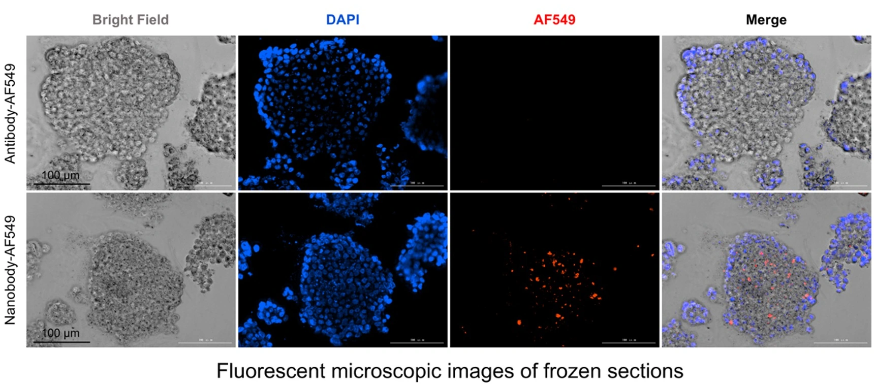
Breast cancer PDO studies combine frozen-section distribution of AF549-labeled antibody/nanobody formats with morphology and viability after ADC treatment. A new candidate program can add target profiling, imaging controls, dedicated penetration quantification and killing endpoints.
 View image
Frozen-section spatial distribution: bright-field, DAPI, AF549 and merged views compare relative signal patterns for labeled antibody and nanobody in PDO frozen sections.
TRiCBIO breast cancer PDO antibody/nanobody distribution study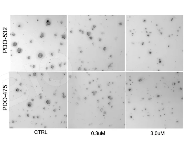 View image
Post-treatment morphology: bright-field views under control, 0.3 μM and 3.0 μM conditions provide tissue context for viability readouts.
TRiCBIO breast cancer PDO ADC study data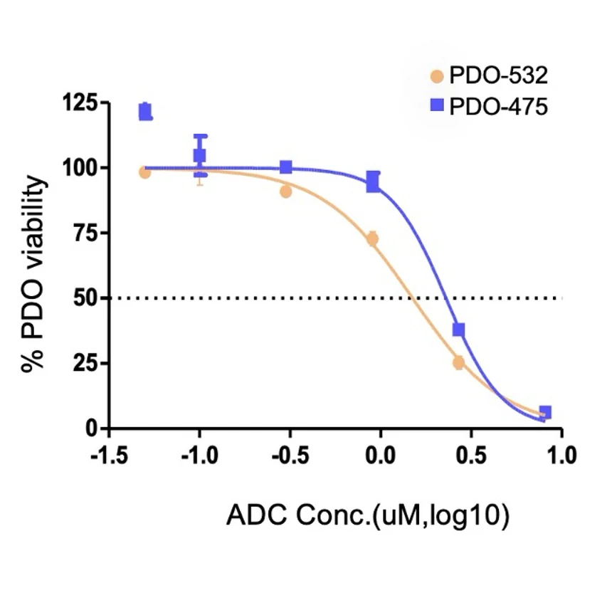 View image
Dose response: two PDO models show different viability profiles across a candidate ADC concentration range, informing replication and follow-on sample studies.
TRiCBIO breast cancer PDO ADC study dataContinue to the related case for model context, experimental setup and interpretation.
Study focus
Study workflow
Use histology, IHC or IF to assess target expression and spatial heterogeneity, and confirm model identity and culture quality.
Define labeling, incubation, imaging and competition or target-negative controls around molecule properties; add dedicated imaging and quantification when intact-organoid penetration is a priority.
Interpret spatial distribution with dose response, viability, apoptosis and histology.
Compare candidates consistently and deliver original images, quantitative analysis and interpretation.
Model options
ADC studies first compare candidates through section imaging and functional readouts; quantitative penetration studies can then add serial sections or z-stacks, distance/intensity profiles, time points and target/competition controls.
PROJECT DISCUSSION
ADC design and target biology determine the tumor models, distribution imaging, killing and bystander endpoints for the study.
Application
Compare ADC candidates in human organoids using target-expression, tissue-distribution and activity readouts, with dedicated studies of bystander killing.
Spatial imaging and activity studies
Breast cancer PDO studies combine frozen-section distribution of AF549-labeled antibody/nanobody formats with morphology and viability after ADC treatment. A new candidate program can add target profiling, imaging controls, dedicated penetration quantification and killing endpoints.
View imageFrozen-section spatial distribution: bright-field, DAPI, AF549 and merged views compare relative signal patterns for labeled antibody and nanobody in PDO frozen sections.
TRiCBIO breast cancer PDO antibody/nanobody distribution studyView imagePost-treatment morphology: bright-field views under control, 0.3 μM and 3.0 μM conditions provide tissue context for viability readouts.
TRiCBIO breast cancer PDO ADC study dataView imageDose response: two PDO models show different viability profiles across a candidate ADC concentration range, informing replication and follow-on sample studies.
TRiCBIO breast cancer PDO ADC study dataContinue to the related case for model context, experimental setup and interpretation.
Study focus
Study workflow
Use histology, IHC or IF to assess target expression and spatial heterogeneity, and confirm model identity and culture quality.
Define labeling, incubation, imaging and competition or target-negative controls around molecule properties; add dedicated imaging and quantification when intact-organoid penetration is a priority.
Interpret spatial distribution with dose response, viability, apoptosis and histology.
Compare candidates consistently and deliver original images, quantitative analysis and interpretation.
Model options
ADC studies first compare candidates through section imaging and functional readouts; quantitative penetration studies can then add serial sections or z-stacks, distance/intensity profiles, time points and target/competition controls.
PROJECT DISCUSSION
ADC design and target biology determine the tumor models, distribution imaging, killing and bystander endpoints for the study.