
Immunotherapy and combinations
Study checkpoint inhibitors, immune combinations and treatment-associated cellular or secreted changes.
Technology platform
Study treatment response in human 3D tumors alongside source-tissue architecture and immune cells retained from the sample, with histology, flow cytometry, cytokine and functional readouts.
Study applications
Study checkpoint inhibitors, immune combinations and treatment-associated cellular or secreted changes.
Profile T, B, NK, macrophage and other immune populations present in the sample.
Relate H&E, IHC or IF findings to flow, cytokine and efficacy results.
Supports studies centered on tumor epithelial phenotype, target expression and standardized drug response.
Brings tissue architecture, the sample’s immune-cell context and treatment response into one study, informing the culture window and assay endpoints.
PDO 2.0 studies & assays
See how TRiCBIO combines tissue morphology, immune profiling and treatment-related flow readouts in ccRCC and NSCLC PDO 2.0 studies.
 View image
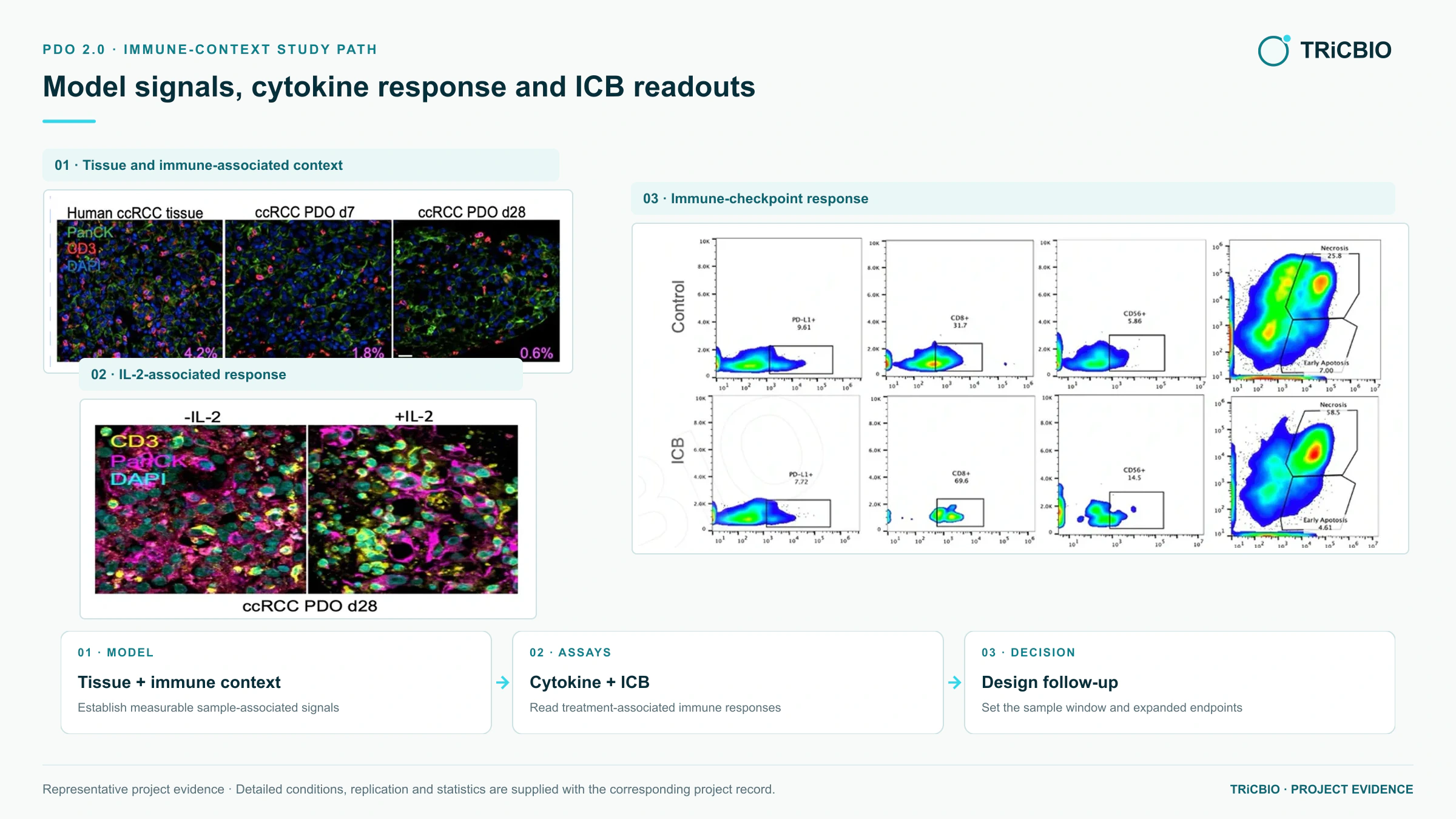
PDO 2.0 TME study: tissue imaging and flow assess immune-associated signals together with early-apoptosis and necrosis changes under treatment conditions, with sample, treatment and gating details included in the study deliverables.
TRiCBIO ccRCC PDO 2.0 immune-context study dataOriginal method and functional evidence
Figure 1 presents the establishment and characterization of ALI patient-derived tumor organoids across tumor types. Figure 7 studies TIL activation, expansion and tumor-cell killing under PD-1-related conditions in selected samples.
TRiCBIO applies the ALI approach to baseline sample profiling, immune-readout selection and study-window design for PDO 2.0 programs.
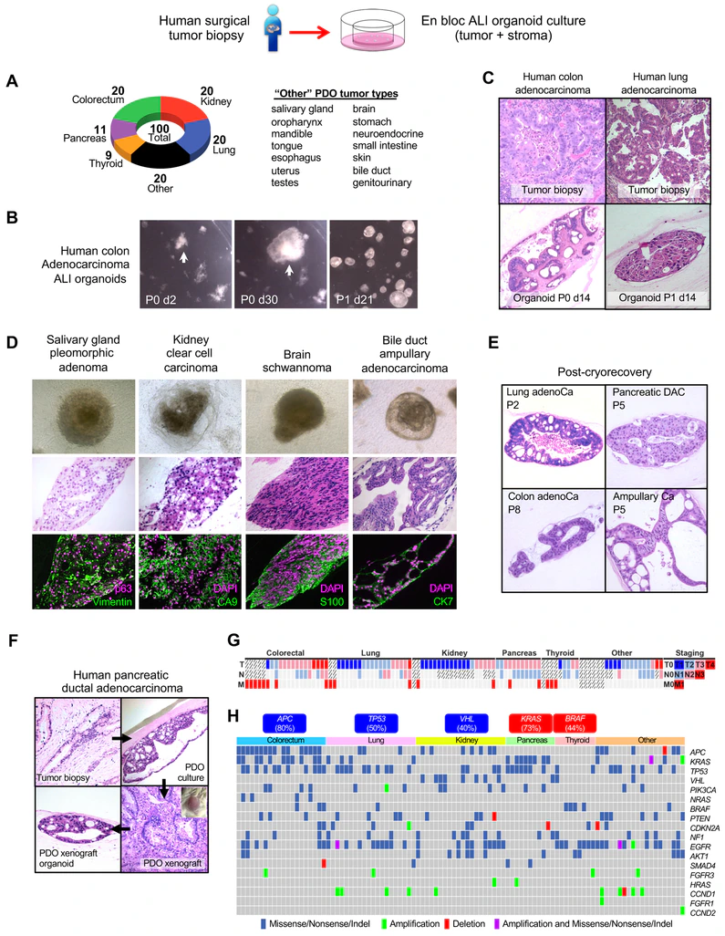
Culture morphology, source-tissue/PDO histology, lineage markers and genomic alterations characterize model establishment and identity.
Neal JT, Li X et al. Cell. 2018;175:1972–1988.e16, Figure 1.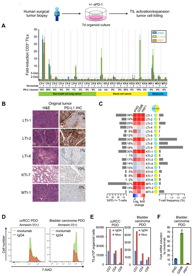
Immune baseline, cytokine signals, TIL counts and tumor-cell killing are combined into a multidimensional functional profile for immunotherapy studies.
Neal JT, Li X et al. Cell. 2018;175:1972–1988.e16, Figure 7.Nature Reviews Cancer introduced the “Organoid 2.0” framing for ALI tumor–immune organoids in 2019 and revisited tumor–immune organoid models and applications in depth in 2024. For immunotherapy, complex drug mechanisms and tissue-level response studies, this direction adds biological context beyond epithelial-only organoids.
The article framed the Cell 2018 ALI tumor–immune organoid study as “Organoid 2.0”: extending the model beyond tumor epithelium to include source-sample tissue architecture and immune context for tumor–immune interaction studies.
DOI 10.1038/s41568-019-0108-x ↗2024 · Review ArticleThe review maps tumor–immune organoid models, immune-analysis approaches and drug-research applications, showing how tissue and immune context can inform the study of treatment response and mechanism.
DOI 10.1038/s41568-024-00706-6 ↗Plan your study
Use the sample baseline, candidate mechanism and study endpoint to define the culture window, controls and assay plan, then organize the results for review and follow-on work.
Tumor type, sample source and condition, ethics documentation, candidate, dosing plan and study objective.
Model establishment and QC, treatment, H&E, IHC, IF, flow cytometry, cytokines and TCR analysis, with functional endpoints selected for the study.
Model QC, methods, raw data, images, statistical analysis, key findings and study report.
Assay endpoints are selected around tissue architecture, the sample’s immune context and candidate mechanism so complementary results can be interpreted within one study.
PROJECT DISCUSSION
Tell us the tumor type, sample status and endpoints you need. Our scientists will assess whether ALI / PDO 2.0 fits and propose the assay panel and study scope.
Technology platform
Study treatment response in human 3D tumors alongside source-tissue architecture and immune cells retained from the sample, with histology, flow cytometry, cytokine and functional readouts.
Study applications
Study checkpoint inhibitors, immune combinations and treatment-associated cellular or secreted changes.
Profile T, B, NK, macrophage and other immune populations present in the sample.
Relate H&E, IHC or IF findings to flow, cytokine and efficacy results.
Supports studies centered on tumor epithelial phenotype, target expression and standardized drug response.
Brings tissue architecture, the sample’s immune-cell context and treatment response into one study, informing the culture window and assay endpoints.
PDO 2.0 studies & assays
See how TRiCBIO combines tissue morphology, immune profiling and treatment-related flow readouts in ccRCC and NSCLC PDO 2.0 studies.
View imagePDO 2.0 TME study: tissue imaging and flow assess immune-associated signals together with early-apoptosis and necrosis changes under treatment conditions, with sample, treatment and gating details included in the study deliverables.
TRiCBIO ccRCC PDO 2.0 immune-context study dataOriginal method and functional evidence
Figure 1 presents the establishment and characterization of ALI patient-derived tumor organoids across tumor types. Figure 7 studies TIL activation, expansion and tumor-cell killing under PD-1-related conditions in selected samples.
TRiCBIO applies the ALI approach to baseline sample profiling, immune-readout selection and study-window design for PDO 2.0 programs.
Culture morphology, source-tissue/PDO histology, lineage markers and genomic alterations characterize model establishment and identity.
Neal JT, Li X et al. Cell. 2018;175:1972–1988.e16, Figure 1.Immune baseline, cytokine signals, TIL counts and tumor-cell killing are combined into a multidimensional functional profile for immunotherapy studies.
Neal JT, Li X et al. Cell. 2018;175:1972–1988.e16, Figure 7.Nature Reviews Cancer introduced the “Organoid 2.0” framing for ALI tumor–immune organoids in 2019 and revisited tumor–immune organoid models and applications in depth in 2024. For immunotherapy, complex drug mechanisms and tissue-level response studies, this direction adds biological context beyond epithelial-only organoids.
The article framed the Cell 2018 ALI tumor–immune organoid study as “Organoid 2.0”: extending the model beyond tumor epithelium to include source-sample tissue architecture and immune context for tumor–immune interaction studies.
DOI 10.1038/s41568-019-0108-x ↗2024 · Review ArticleThe review maps tumor–immune organoid models, immune-analysis approaches and drug-research applications, showing how tissue and immune context can inform the study of treatment response and mechanism.
DOI 10.1038/s41568-024-00706-6 ↗Plan your study
Use the sample baseline, candidate mechanism and study endpoint to define the culture window, controls and assay plan, then organize the results for review and follow-on work.
Tumor type, sample source and condition, ethics documentation, candidate, dosing plan and study objective.
Model establishment and QC, treatment, H&E, IHC, IF, flow cytometry, cytokines and TCR analysis, with functional endpoints selected for the study.
Model QC, methods, raw data, images, statistical analysis, key findings and study report.
Assay endpoints are selected around tissue architecture, the sample’s immune context and candidate mechanism so complementary results can be interpreted within one study.
PROJECT DISCUSSION
Tell us the tumor type, sample status and endpoints you need. Our scientists will assess whether ALI / PDO 2.0 fits and propose the assay panel and study scope.