
Advanced custom co-development
Vascular phenotypes & co-culture
Profile vascular phenotypes and spatial organization, with endothelial or mural-cell co-culture developed when required by the study.
Histology and spatial phenotype
Vascular-associated phenotypes in tissue and PDO
Glioma, HCC and ALI organoid studies bring together tissue morphology, vascular markers and spatial co-localization, providing a strong starting point for vascular-context, co-culture and functional studies.
Tissue–PDO comparisonH&E · CD31 · HEP-1Spatial co-localization
View the complete glioma PDO 2.0 case ↗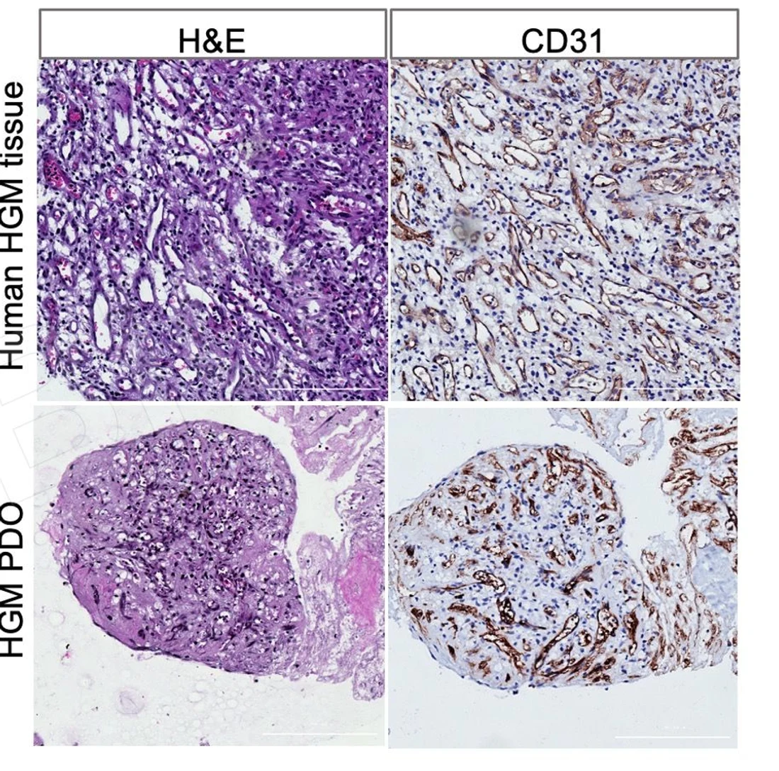 View image
Top: source tissue; bottom: PDO. H&E and CD31 staining show tissue morphology and CD31 staining patterns in each.
TRiCBIO glioma PDO 2.0 study data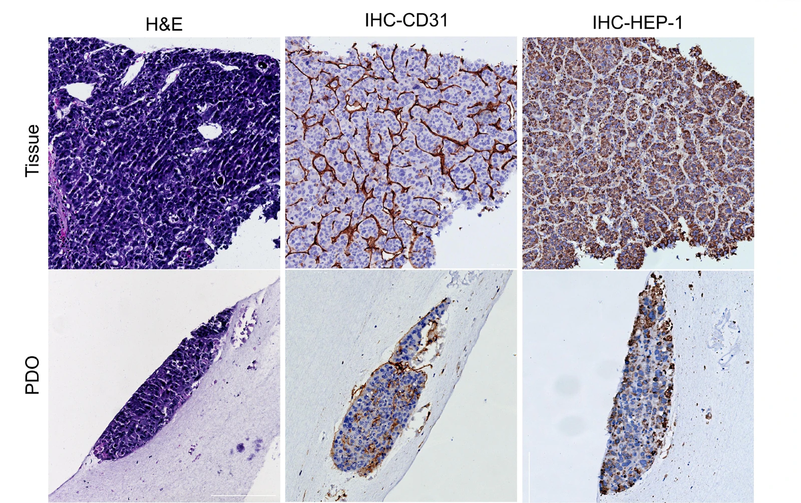 View image
Matched H&E, CD31 and HEP-1 panels connect tissue identity with vascular features in the source tissue and PDO.
TRiCBIO HCC organoid study data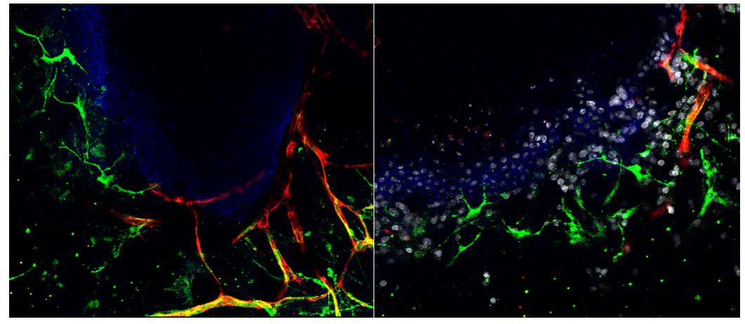 View image
Joint imaging of CD31, PGP9.5 and E-cadherin visualizes spatial relationships among vascular-, neural- and epithelial-associated signals.
TRiCBIO ALI organoid spatial imaging dataStudy applications
Where vascular-focused co-culture adds value
Observe vascular markers, spatial organization and tissue interaction
Develop complex co-culture for vascular-context studies
Custom-development process
From cellular composition and spatial structure to functional studies
Start with cell composition and co-culture compatibility, then combine structural markers and functional endpoints into a model and assay strategy for vascular mechanisms, dosing or safety studies.
Cell system and study objective
Identify the endothelial, mural or other cellular components needed and the interaction the study should resolve.
Co-culture design and baseline profiling
Assess survival, spatial distribution, culture compatibility and baseline structure.
Structural and spatial profiling
Use CD31 / PECAM-1, α-SMA and imaging endpoints to characterize cell identity, spatial organization and tissue interaction.
Intervention results and next step
Use the study findings to progress into dosing, toxicology or mechanism work.
Deliverables
Use study results to plan follow-on development
- Co-culture performance and morphology
- Cell identity, spatial distribution and structural QC
- Study report and recommendations for follow-on development
Related models
Select the starting model by tissue context and endpoint
Complex co-culture studies begin with vascular markers, cell composition, culture compatibility and spatial interactions, then add the structural and functional endpoints required by the program.
PROJECT DISCUSSION
Discuss vascular-focused co-culture or custom development
Share the target tissue, cellular components and priority structural or functional endpoints. We’ll assess co-culture compatibility, markers and the most suitable study design.