01Histology and lineage
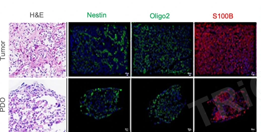
Source-tissue and PDO histology and markers
H&E, Nestin, Olig2 and S100B place tumor-tissue and PDO lineage context side by side.
Establishes the model-identity baseline for treatment and mechanism studiesExplore glioma PDO 2.0 through histology, lineage markers, CD31 and treatment-response studies.
STUDY EVIDENCE
Complementary glioma PDO 2.0 studies separately show model identity and lineage profiling, CD31-associated context and treatment-response readouts.
H&E, Nestin, Olig2 and S100B place tumor-tissue and PDO lineage context side by side.
Establishes the model-identity baseline for treatment and mechanism studies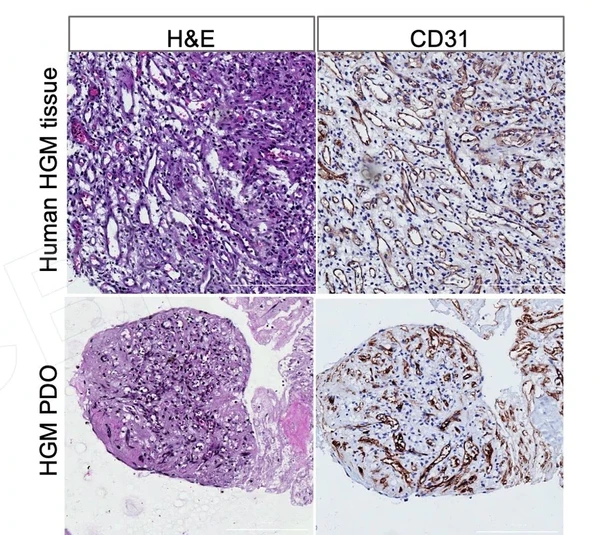
A complementary glioma study extends the vascular context through tissue/PDO morphology and CD31-associated profiling.
Helps define spatial, vascular and follow-on mechanism endpoints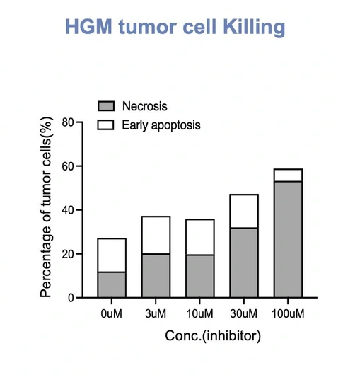
A complementary study uses early-apoptosis and necrosis readouts to show functional changes across treatment concentrations.
Informs mechanism studies, replication and work in additional samplesINTERPRETATION
Helps define model-identity, vascular, treatment and mechanism endpoints.
Histology, lineage, vascular and treatment-response readouts support a multidimensional glioma PDO 2.0 study design, with endpoints selected for the sample and candidate.
Add dose and time-course studies informed by molecular context, followed by mechanism endpoints and independent-model validation.
Related models and services
PROJECT DISCUSSION
Tell us which model, assay or finding you want to extend. Our scientists will adapt the approach to your candidate, samples and decision point.
Explore glioma PDO 2.0 through histology, lineage markers, CD31 and treatment-response studies.
STUDY EVIDENCE
Complementary glioma PDO 2.0 studies separately show model identity and lineage profiling, CD31-associated context and treatment-response readouts.
H&E, Nestin, Olig2 and S100B place tumor-tissue and PDO lineage context side by side.
Establishes the model-identity baseline for treatment and mechanism studiesA complementary glioma study extends the vascular context through tissue/PDO morphology and CD31-associated profiling.
Helps define spatial, vascular and follow-on mechanism endpointsA complementary study uses early-apoptosis and necrosis readouts to show functional changes across treatment concentrations.
Informs mechanism studies, replication and work in additional samplesINTERPRETATION
Helps define model-identity, vascular, treatment and mechanism endpoints.
Histology, lineage, vascular and treatment-response readouts support a multidimensional glioma PDO 2.0 study design, with endpoints selected for the sample and candidate.
Add dose and time-course studies informed by molecular context, followed by mechanism endpoints and independent-model validation.
Related models and services
PROJECT DISCUSSION
Tell us which model, assay or finding you want to extend. Our scientists will adapt the approach to your candidate, samples and decision point.