
012D cell system
Fast, controlled early comparison
Initial target or pathway screening
Simplified biological context
Compare 2D, organoid and PDX models by biological context, timing and throughput to choose the system that best fits the program goal.
MODEL SELECTION MAP
Select or combine models around the study objective, biological context, scale and timing.
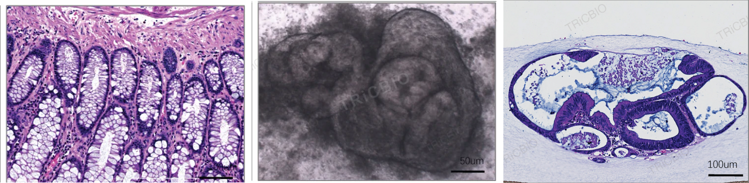
The three panels show source-tissue H&E, PDO bright-field morphology and PDO histology, comparing culture morphology with the original tissue.
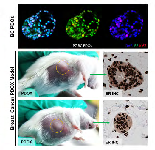
Model & assay dataIdentity from in vitro to in vivo: ER and Ki67 staining in a breast cancer PDO is shown alongside ER immunohistochemistry in PDOX tissue to compare receptor phenotype and model identity.
Study dataConnect human in vitro model characterization with PDOX-based in vivo studies, choosing each step around the candidate and endpoint.
Fast, controlled early comparison
Initial target or pathway screening
Simplified biological context
Provides 3D tissue phenotypes and donor-derived context
Candidate, tissue-response, mechanism and safety studies
Match model complexity to sample conditions
Extends organoid findings into an in vivo setting
Exposure, in vivo response and translational extension
Time, resources and in vivo study design
COMMON SEQUENCE
Use 2D assays for broad early screening, then bring selected candidates into organoid models when tissue architecture, cellular interactions or human tissue context can sharpen the decision.
Organoids balance human origin, 3D structure, sample variation and experimental scale for efficacy, mechanism and safety questions closer to the tissue level.
In vivo models extend selected findings into growth, histology and pharmacology after the relevant in vitro signal and model identity are established.
Use each system for the evidence it contributes, then combine rapid comparison, human tissue context and in vivo extension around the program objective.
Related models and services
PROJECT DISCUSSION
Share your project goal and available materials. Our scientists will recommend models, controls, assays and a practical first study.
Compare 2D, organoid and PDX models by biological context, timing and throughput to choose the system that best fits the program goal.
MODEL SELECTION MAP
Select or combine models around the study objective, biological context, scale and timing.
The three panels show source-tissue H&E, PDO bright-field morphology and PDO histology, comparing culture morphology with the original tissue.
Model & assay dataIdentity from in vitro to in vivo: ER and Ki67 staining in a breast cancer PDO is shown alongside ER immunohistochemistry in PDOX tissue to compare receptor phenotype and model identity.
Study dataConnect human in vitro model characterization with PDOX-based in vivo studies, choosing each step around the candidate and endpoint.
Fast, controlled early comparison
Initial target or pathway screening
Simplified biological context
Provides 3D tissue phenotypes and donor-derived context
Candidate, tissue-response, mechanism and safety studies
Match model complexity to sample conditions
Extends organoid findings into an in vivo setting
Exposure, in vivo response and translational extension
Time, resources and in vivo study design
COMMON SEQUENCE
Use 2D assays for broad early screening, then bring selected candidates into organoid models when tissue architecture, cellular interactions or human tissue context can sharpen the decision.
Organoids balance human origin, 3D structure, sample variation and experimental scale for efficacy, mechanism and safety questions closer to the tissue level.
In vivo models extend selected findings into growth, histology and pharmacology after the relevant in vitro signal and model identity are established.
Use each system for the evidence it contributes, then combine rapid comparison, human tissue context and in vivo extension around the program objective.
Related models and services
PROJECT DISCUSSION
Share your project goal and available materials. Our scientists will recommend models, controls, assays and a practical first study.