
Objective and research priorities
Review sample source, disease or tissue features, existing methods and the intended question to identify the one or two endpoints that matter most.
R&D service
Develop a model and assay plan for specialized tissues, rare diseases or complex co-cultures, starting from the samples and endpoints available to your program.
 View image
Custom model studies
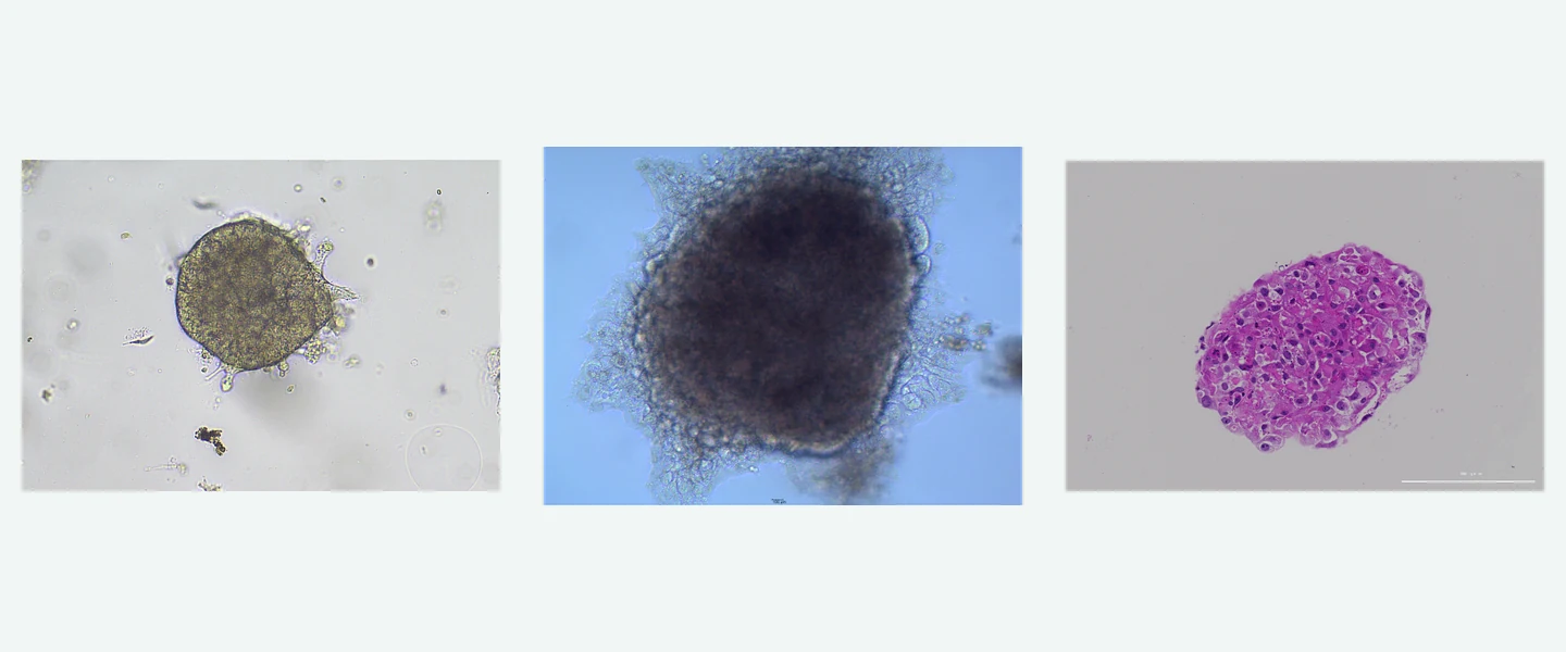
Head and neck cancer culture and histology, adipose differentiation with Oil Red O, and pathology-guided GIST and osteosarcoma studies show how specialized models begin with sample context and identity QC, then progress into functional and candidate-response studies.
Study focus
Study workflow
Custom development supports specialized tissues, rare diseases, complex co-cultures and new assays.
Current project experience includes head and neck cancer organoid culture and histology, adipose differentiation and lipid accumulation, and pathology-guided mesenchymal tumor models. We design culture and assays around sample conditions, key phenotypes and intended use.
Review sample source, disease or tissue features, existing methods and the intended question to identify the one or two endpoints that matter most.
Configure samples and conditions around culture, cellular composition, key phenotype and assay window, creating a clear path to model expansion and follow-on applications.
Use prespecified criteria to plan follow-on work and optimize culture, controls, repeatability and assay methods.
Use project findings to expand samples or batches and document scope, SOP, QC criteria, raw data and recommendations for future use.
Study images and readouts
Head and neck cancer culture morphology and H&E, adipose phenotypes before and after differentiation, and source-tissue/model comparisons in mesenchymal tumors show how specialized models are established, checked against source tissue and prepared for functional readouts. Efficacy, mechanism or co-culture studies can then be added for the program.
 View image
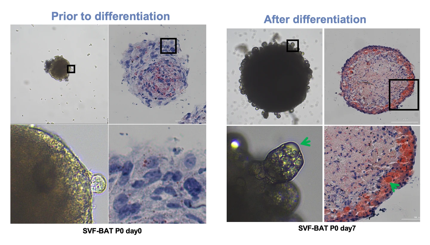
Adipose custom model: bright-field morphology and Oil Red O staining show a lipid-accumulation phenotype before and after differentiation, providing a measurable starting point for metabolic biology, lipid handling and candidate-response studies.
Study data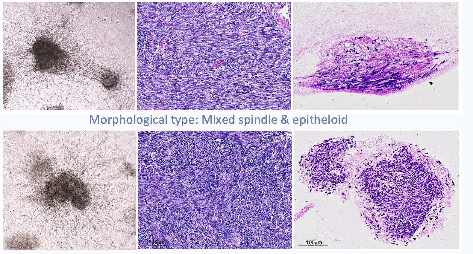 View image
Custom GIST models: bright-field culture morphology, source-tissue H&E and culture H&E compare spindle and mixed spindle/epithelioid patterns for case-specific pathology reference and model QC.
Study dataView the mesenchymal-tumor study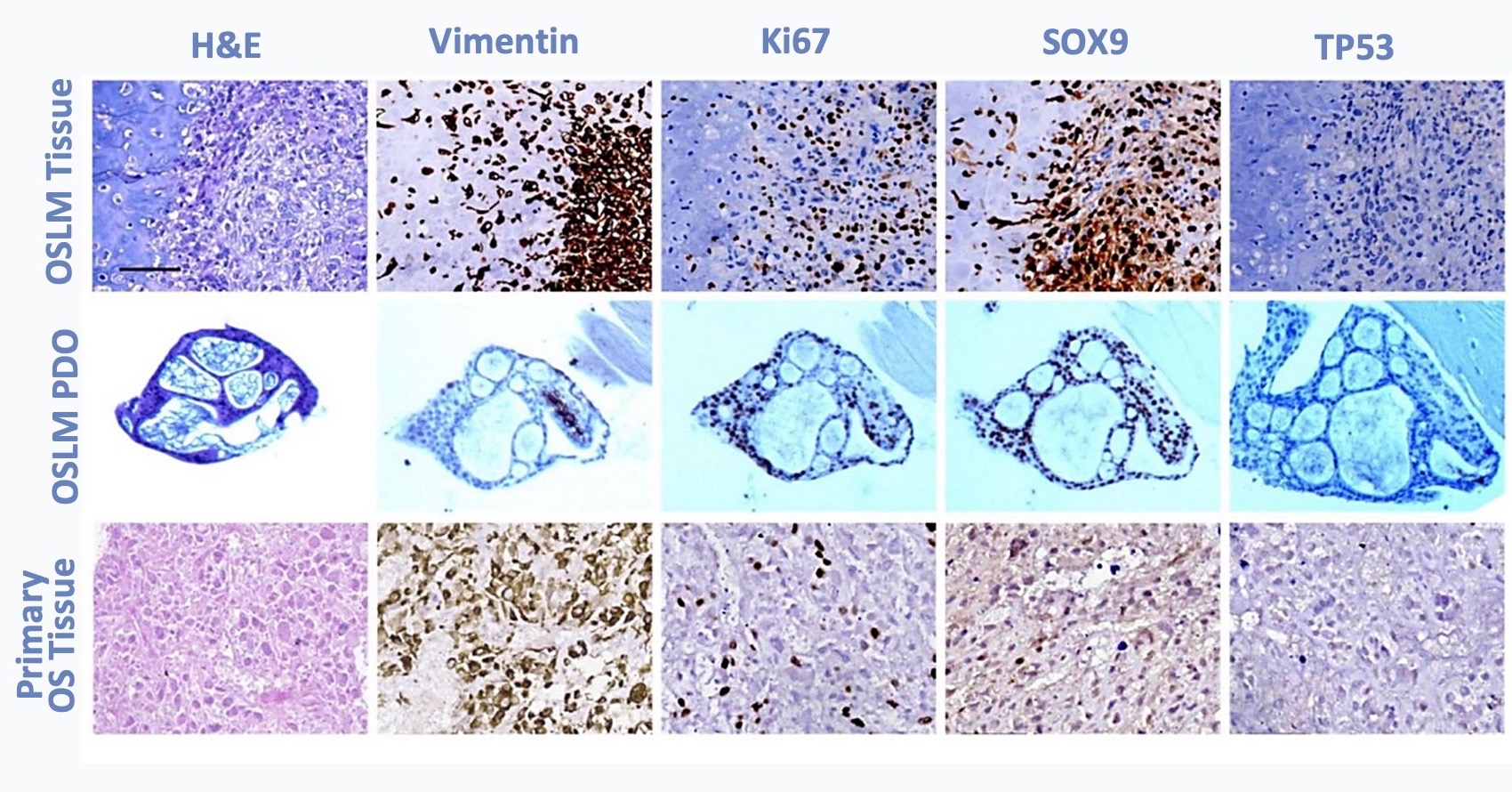 View image
Custom osteosarcoma models: H&E and marker panels across source tissue, PDO and primary tissue compare identity, proliferation and lineage-associated phenotypes for downstream assay selection.
Study dataView the mesenchymal-tumor studyExpected study outputs
Experimental conditions, original images or data, QC results and an explanation of the findings.
A clear summary of what the model can assess, available assay options and the factors that affect model establishment and reproducibility.
SOPs, QC guidance and a method report, together with a plan for the next study.
Relevant model systems
Programs can start from an established model or new samples, with a clear development path built around source material, primary endpoints and critical controls before extending into efficacy, mechanism, safety or method transfer.
For BBB, ADPKD and complex co-cultures, TRiCBIO builds from kidney organoid data, HUVEC–astrocyte co-culture, tight-junction imaging and permeability methods, then adds the disease phenotype, cell source and functional readouts the program requires.
PROJECT DISCUSSION
A tissue or disease context, sample source and key phenotype or assay need are enough to begin. Together we can plan model establishment, QC and follow-on studies.
R&D service
Develop a model and assay plan for specialized tissues, rare diseases or complex co-cultures, starting from the samples and endpoints available to your program.
View imageCustom model studies
Head and neck cancer culture and histology, adipose differentiation with Oil Red O, and pathology-guided GIST and osteosarcoma studies show how specialized models begin with sample context and identity QC, then progress into functional and candidate-response studies.
Study focus
Study workflow
Custom development supports specialized tissues, rare diseases, complex co-cultures and new assays.
Current project experience includes head and neck cancer organoid culture and histology, adipose differentiation and lipid accumulation, and pathology-guided mesenchymal tumor models. We design culture and assays around sample conditions, key phenotypes and intended use.
Review sample source, disease or tissue features, existing methods and the intended question to identify the one or two endpoints that matter most.
Configure samples and conditions around culture, cellular composition, key phenotype and assay window, creating a clear path to model expansion and follow-on applications.
Use prespecified criteria to plan follow-on work and optimize culture, controls, repeatability and assay methods.
Use project findings to expand samples or batches and document scope, SOP, QC criteria, raw data and recommendations for future use.
Study images and readouts
Head and neck cancer culture morphology and H&E, adipose phenotypes before and after differentiation, and source-tissue/model comparisons in mesenchymal tumors show how specialized models are established, checked against source tissue and prepared for functional readouts. Efficacy, mechanism or co-culture studies can then be added for the program.
View imageAdipose custom model: bright-field morphology and Oil Red O staining show a lipid-accumulation phenotype before and after differentiation, providing a measurable starting point for metabolic biology, lipid handling and candidate-response studies.
Study dataView imageCustom GIST models: bright-field culture morphology, source-tissue H&E and culture H&E compare spindle and mixed spindle/epithelioid patterns for case-specific pathology reference and model QC.
Study dataView the mesenchymal-tumor studyView imageCustom osteosarcoma models: H&E and marker panels across source tissue, PDO and primary tissue compare identity, proliferation and lineage-associated phenotypes for downstream assay selection.
Study dataView the mesenchymal-tumor studyExpected study outputs
Experimental conditions, original images or data, QC results and an explanation of the findings.
A clear summary of what the model can assess, available assay options and the factors that affect model establishment and reproducibility.
SOPs, QC guidance and a method report, together with a plan for the next study.
Relevant model systems
Programs can start from an established model or new samples, with a clear development path built around source material, primary endpoints and critical controls before extending into efficacy, mechanism, safety or method transfer.
For BBB, ADPKD and complex co-cultures, TRiCBIO builds from kidney organoid data, HUVEC–astrocyte co-culture, tight-junction imaging and permeability methods, then adds the disease phenotype, cell source and functional readouts the program requires.
PROJECT DISCUSSION
A tissue or disease context, sample source and key phenotype or assay need are enough to begin. Together we can plan model establishment, QC and follow-on studies.