
Barrier system and study objective
Identify the relevant interface—intestinal epithelium, blood–brain barrier or another tissue system—and whether the priority is transport, retention, injury or repair.
R&D service
Assess barrier integrity, injury and repair, then develop permeability or candidate-transport assays for the relevant tissue system.
 View image
Intestinal barrier evidence
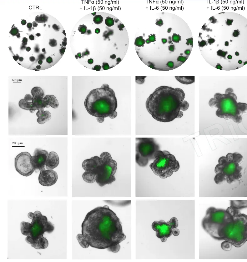
Mouse intestinal organoid studies combine FITC-Dextran permeability with F-actin/ZO-1 epithelial-junction readouts. Human IBD and BBB studies adapt the cell system, identity markers, tight-junction imaging and permeability assays to the available sample and study objective.
Study focus
Study workflow
Confirmed barrier identity, baseline function and assay performance give permeability results a clear biological context. The study brings together tissue-barrier context, delivery format, transport readouts and potential barrier injury.
Identify the relevant interface—intestinal epithelium, blood–brain barrier or another tissue system—and whether the priority is transport, retention, injury or repair.
Confirm cell identity, architecture, tight junctions and baseline permeability against predefined acceptance criteria before candidate testing.
Set donor/receiver orientation, time, concentration and controls around modality, formulation and exposure route, using FITC-Dextran, tracing, imaging or project-appropriate quantification.
Analyze permeability with viability, morphology, F-actin, ZO-1 and other junction readouts to build an integrated view of candidate transport and barrier status.
Study images and readouts
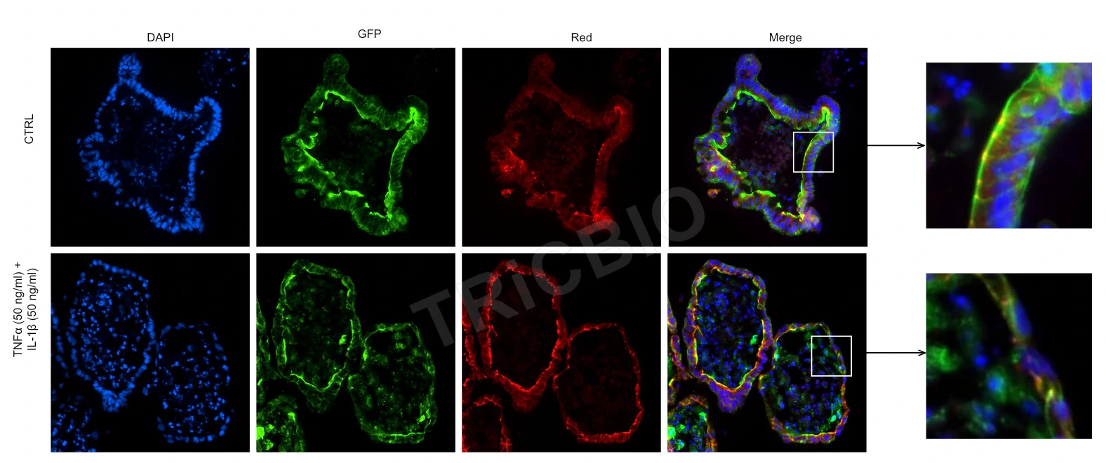
FITC-Dextran, F-actin and ZO-1 results connect permeability change with epithelial structure for candidate and formulation studies.
 View image
Cytoskeleton and tight junctions: F-actin and ZO-1 immunofluorescence provides epithelial-structure and junction-status readouts.
TRiCBIO mouse intestinal organoid studyView the IBD model and barrier studyExpected study outputs
Model identity, tight-junction and baseline permeability results, with batch QC criteria.
Candidate transport or retention data alongside changes in viability, structure and barrier integrity.
Methods, raw data, study conditions and recommended next experiments, including recovery or mass-balance results where applicable.
Relevant model systems
TRiCBIO intestinal organoid studies combine morphology, permeability, viability and F-actin/ZO-1 readouts, with human programs designed around the sample and candidate. Blood–brain barrier studies extend this approach through endothelial–astrocyte systems, tight-junction imaging, and readouts of permeability and candidate effects.
PROJECT DISCUSSION
The candidate or formulation, target barrier, exposure conditions and permeability question shape the model, controls and assay panel.
R&D service
Assess barrier integrity, injury and repair, then develop permeability or candidate-transport assays for the relevant tissue system.
View imageIntestinal barrier evidence
Mouse intestinal organoid studies combine FITC-Dextran permeability with F-actin/ZO-1 epithelial-junction readouts. Human IBD and BBB studies adapt the cell system, identity markers, tight-junction imaging and permeability assays to the available sample and study objective.
Study focus
Study workflow
Confirmed barrier identity, baseline function and assay performance give permeability results a clear biological context. The study brings together tissue-barrier context, delivery format, transport readouts and potential barrier injury.
Identify the relevant interface—intestinal epithelium, blood–brain barrier or another tissue system—and whether the priority is transport, retention, injury or repair.
Confirm cell identity, architecture, tight junctions and baseline permeability against predefined acceptance criteria before candidate testing.
Set donor/receiver orientation, time, concentration and controls around modality, formulation and exposure route, using FITC-Dextran, tracing, imaging or project-appropriate quantification.
Analyze permeability with viability, morphology, F-actin, ZO-1 and other junction readouts to build an integrated view of candidate transport and barrier status.
Study images and readouts
FITC-Dextran, F-actin and ZO-1 results connect permeability change with epithelial structure for candidate and formulation studies.
View imageCytoskeleton and tight junctions: F-actin and ZO-1 immunofluorescence provides epithelial-structure and junction-status readouts.
TRiCBIO mouse intestinal organoid studyView the IBD model and barrier studyExpected study outputs
Model identity, tight-junction and baseline permeability results, with batch QC criteria.
Candidate transport or retention data alongside changes in viability, structure and barrier integrity.
Methods, raw data, study conditions and recommended next experiments, including recovery or mass-balance results where applicable.
Relevant model systems
TRiCBIO intestinal organoid studies combine morphology, permeability, viability and F-actin/ZO-1 readouts, with human programs designed around the sample and candidate. Blood–brain barrier studies extend this approach through endothelial–astrocyte systems, tight-junction imaging, and readouts of permeability and candidate effects.
PROJECT DISCUSSION
The candidate or formulation, target barrier, exposure conditions and permeability question shape the model, controls and assay panel.