Source-tissue H&E, organoid bright-field and organoid histology provide a pathology-informed basis for QC and custom culture design in mesenchymal tumors.
01Study objective02Model & design03Assays & findings04Decision & next step
Model & sampleHuman mesenchymal-tumor 3D culture model
ObjectiveConfirm culture state and program-relevant phenotype, then design the custom study
Study conditionsCulture and candidate-treatment conditions selected for the study
AssaysBright-field, morphology records and program-relevant phenotypic assays
Study evidenceTRiCBIO study data
DeliverablesReceive images, assay results and an analysis summary; the report documents the study design, sample size and statistical methods
STUDY EVIDENCE
GIST morphology and osteosarcoma marker profiling
Two mesenchymal-tumor studies show how culture morphology, source pathology and model markers can be combined for identity, quality assessment and downstream assay design.
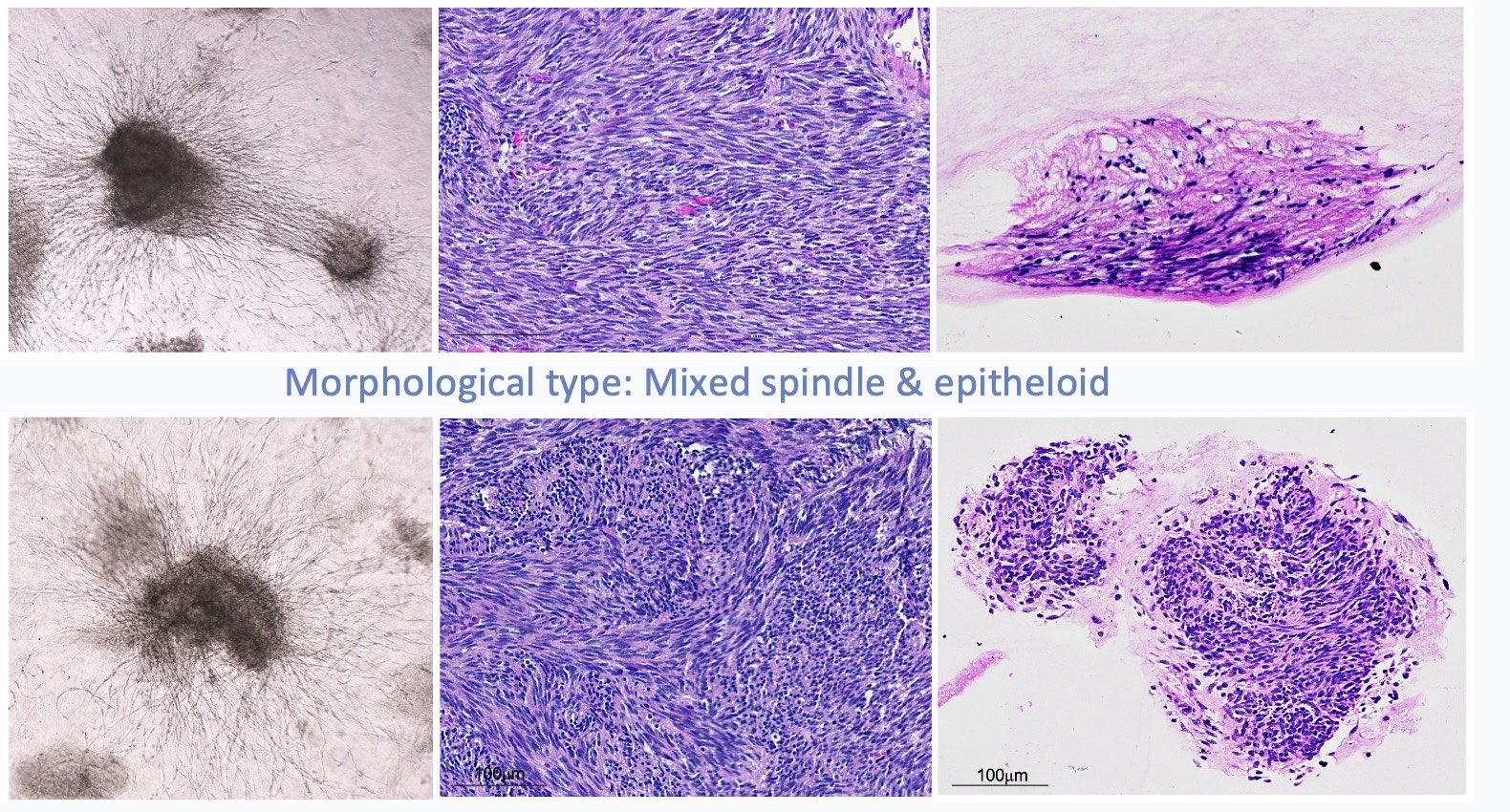
Culture morphology with source-tissue and culture H&E
Bright-field culture morphology, source-tissue H&E and culture H&E compare spindle and mixed spindle/epithelioid patterns within a case-specific pathology reference.
Defines model-QC priorities and downstream identity assays
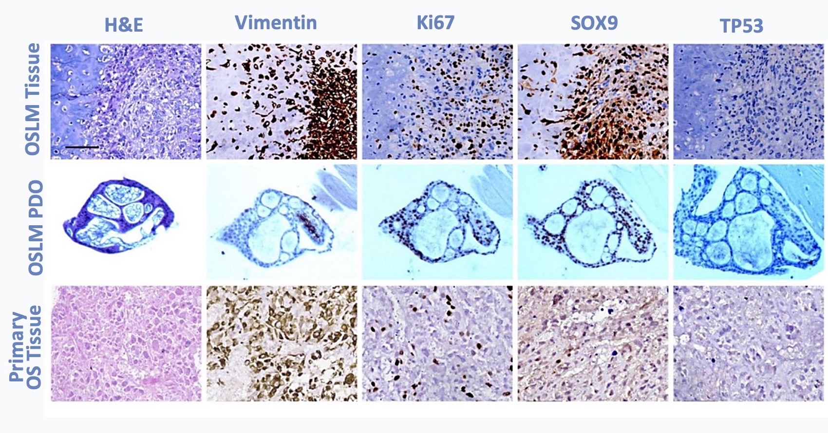
Multidimensional profiling across source tissue, PDO and primary tissue
H&E, Vimentin, Ki67, SOX9 and TP53 panels compare model identity, proliferation and lineage-associated phenotypes.
Guides program-relevant marker and functional endpoint selection
INTERPRETATION
How results guide the next study
How the results are used
Helps select candidate- or mechanism-focused studies suited to the culture system.
Study application
Culture phenotype is interpreted with tissue source, pathology and program-specific QC, then validated through functional endpoints and independent batches.
Follow-on study
Start with source context and appropriate controls, then add dose, time and functional endpoints with independent-batch validation.
Related models and services
Choose the right models and services for the next study
Source-tissue H&E, organoid bright-field and organoid histology provide a pathology-informed basis for QC and custom culture design in mesenchymal tumors.
01Study objective02Model & design03Assays & findings04Decision & next step
Model & sampleHuman mesenchymal-tumor 3D culture model
ObjectiveConfirm culture state and program-relevant phenotype, then design the custom study
Study conditionsCulture and candidate-treatment conditions selected for the study
AssaysBright-field, morphology records and program-relevant phenotypic assays
Study evidenceTRiCBIO study data
DeliverablesReceive images, assay results and an analysis summary; the report documents the study design, sample size and statistical methods
STUDY EVIDENCE
GIST morphology and osteosarcoma marker profiling
Two mesenchymal-tumor studies show how culture morphology, source pathology and model markers can be combined for identity, quality assessment and downstream assay design.
Culture morphology with source-tissue and culture H&E
Bright-field culture morphology, source-tissue H&E and culture H&E compare spindle and mixed spindle/epithelioid patterns within a case-specific pathology reference.
Defines model-QC priorities and downstream identity assays
Multidimensional profiling across source tissue, PDO and primary tissue
H&E, Vimentin, Ki67, SOX9 and TP53 panels compare model identity, proliferation and lineage-associated phenotypes.
Guides program-relevant marker and functional endpoint selection
INTERPRETATION
How results guide the next study
How the results are used
Helps select candidate- or mechanism-focused studies suited to the culture system.
Study application
Culture phenotype is interpreted with tissue source, pathology and program-specific QC, then validated through functional endpoints and independent batches.
Follow-on study
Start with source context and appropriate controls, then add dose, time and functional endpoints with independent-batch validation.
Related models and services
Choose the right models and services for the next study