
PDOX: organoid-to-in vivo validation
Define when a PDO program should extend into PDOX and how model identity, in vivo growth, histology and efficacy are evaluated together.
PDO–PDOX COMPARISON
Compare model identity before extending in vivo
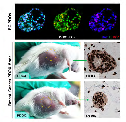
ER profiling in breast PDO/PDOX and MUC5AC staining in gastric PDO/PDOX show how identity markers can be compared across in vitro and in vivo models. Follow-on studies can add tumor growth, efficacy and mechanism endpoints.
 View image
Identity from in vitro to in vivo: ER and Ki67 staining in a breast cancer PDO is shown alongside ER immunohistochemistry in PDOX tissue to compare receptor phenotype and model identity.
Study data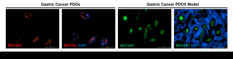 View image
In vitro/in vivo phenotype in a gastric cancer model: MUC5AC immunofluorescence compares marker distribution in PDO and PDOX and provides a phenotype baseline for in vivo extension.
Study dataWhen to use PDOX
Once an in vitro PDO has established identity and a clear response signal, PDOX extends the study into in vivo growth, histology, efficacy and mechanism endpoints.
Confirm the in vitro model first
Check PDO source, passage, histology, target markers, culture state and existing efficacy before animal work.
Compare in vitro and in vivo evidence
Growth, histology, IHC and treatment outcomes can be compared with PDO marker and response trends.
- PDO entry QC
- In vivo growth monitoring
- H&E/IHC identity
- Efficacy and mechanism endpoints
Designing the animal study
Strain, group size, cell input, dosing and duration are selected around the scientific question, ethics approval and statistical plan.
Related models and services
Find relevant models and services
PROJECT DISCUSSION
Discuss your study with a scientist
Share your project goal and available materials. Our scientists will recommend models, controls, assays and a practical first study.