
Normal gastric organoids: culture & histology
P0–P2 culture, H&E, Ki67 and mucin-associated immunofluorescence show growth across passages and epithelial features.
CULTURE, HISTOLOGY & PHENOTYPE
Growth and secretory phenotype
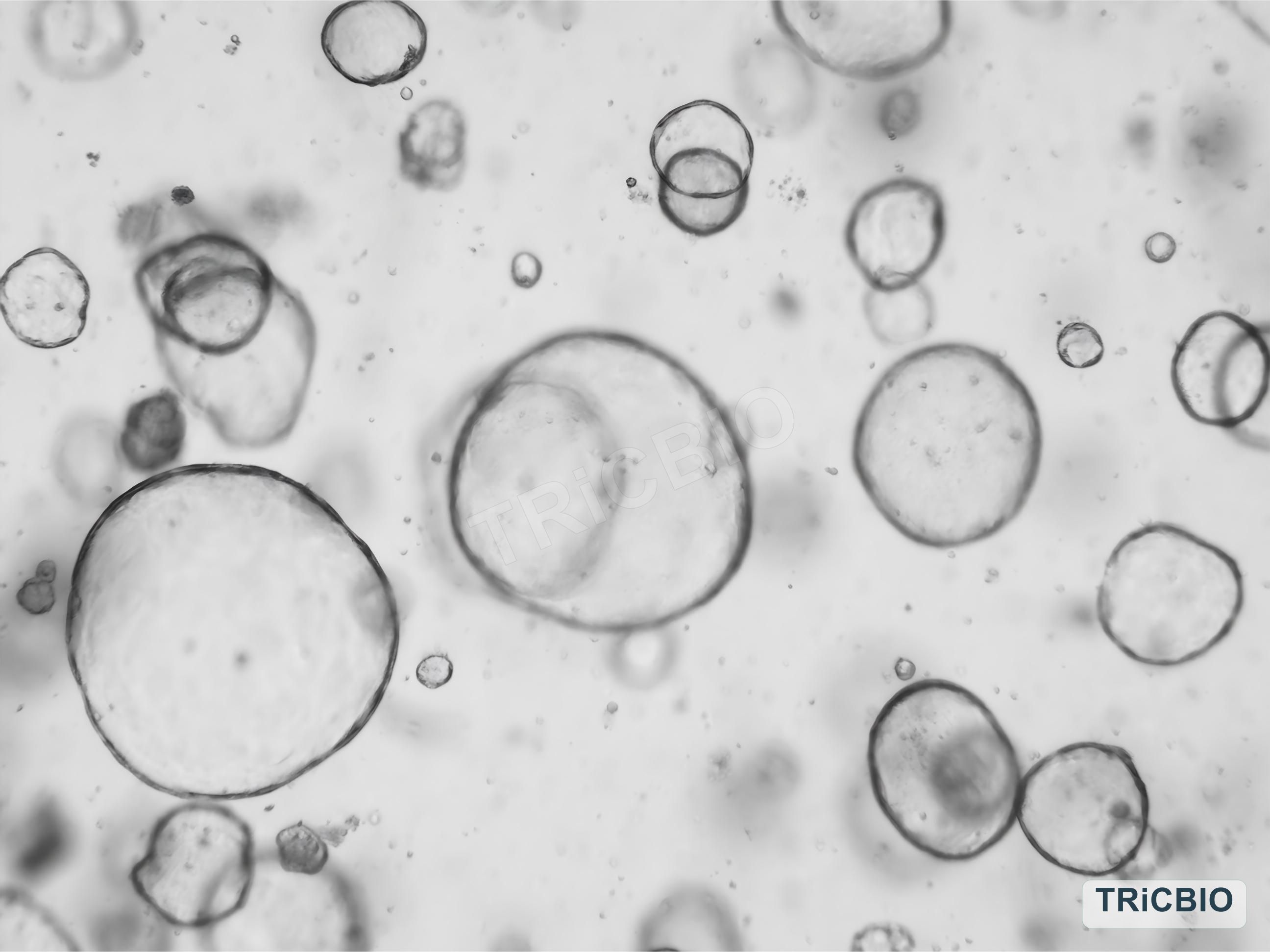
Bright-field imaging and H&E show normal gastric organoid growth and epithelial structure, while Ki67 and mucin-associated immunofluorescence add proliferative and secretory phenotypes.
 View image
Culture morphology: a bright-field image shows normal gastric organoid growth in 3D culture.
Model & assay data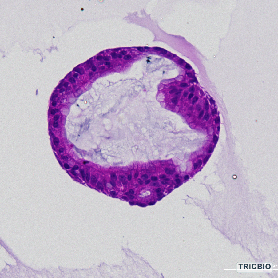 View image
Histology: H&E is used to evaluate epithelial organization and lumen-like structure.
Model & assay data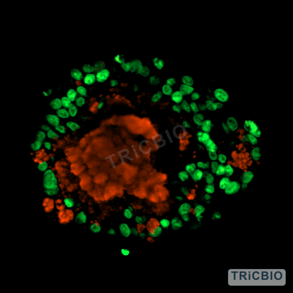 View image
Immunofluorescence: Ki67 and mucin-associated signals add proliferation and secretory context.
Model & assay data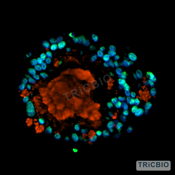 View image
Additional field: proliferation and mucin signals across organoid structures are interpreted with culture, passaging and histology results to assess model status.
Model & assay dataCulture and serial passaging
Serial imaging follows normal gastric organoids from P0 through P2, showing growth and morphology across passages.
Histology and epithelial features
H&E describes tissue architecture, while Ki67 and mucin-associated immunofluorescence add proliferation and secretory context.
- P0–P2 bright-field imaging
- H&E
- Ki67
- Mucin-associated immunofluorescence
Potential study use
Use the model to study gastric epithelial biology, drug exposure and tissue injury with endpoints defined for the program.
Configure gastric epithelial and injury studies
Culture morphology, histology and epithelial features provide the starting point. Batch context, exposure conditions and additional readouts can then be selected for the study objective.
Related models and services
Find relevant models and services
PROJECT DISCUSSION
Discuss your study with a scientist
Tell us which model features and readouts matter most. Our scientists will recommend the model, QC plan and study endpoints.