
Sample and tissue context
Sample source, tissue context, cycle or stage information, and available ethics or background documentation.
Track morphology and marker expression from primary establishment and passaging through proliferative, secretory and receptive stages, with epithelial-identity profiling throughout.
Human endometrial tissue, with sample context, cycle stage and applicable ethics requirements incorporated into the study design
3D culture · Stage-directed differentiation
TRiCBIO human endometrial organoid establishment, passage and stage-characterization data
Model characterization
The model dossier covers establishment, passaging, H&E, epithelial identity and stage-related markers, supporting tissue-biology, stage-differentiation and candidate-response studies.
 View image
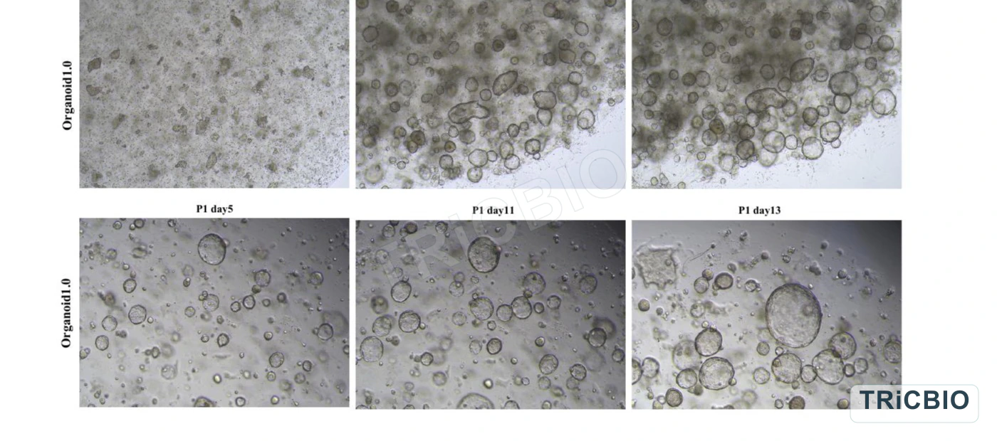
Establishment and passage: the displayed time points document organoid formation after primary processing and continued growth after passage.
TRiCBIO human endometrial organoid characterization data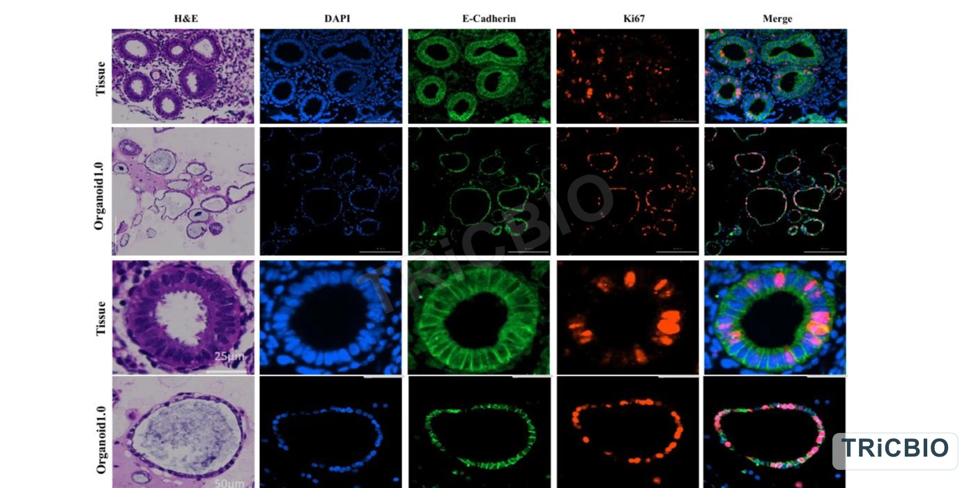 View image
Baseline identity: H&E, DAPI, E-cadherin and Ki67 images compare source tissue and organoids for epithelial organization and proliferative state.
TRiCBIO human endometrial organoid characterization data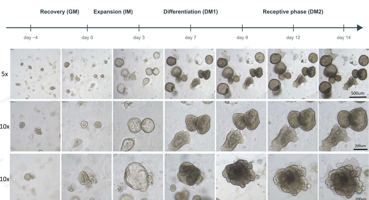 View image
Stage-dependent morphology: serial imaging within one culture workflow helps define time points for downstream assays.
TRiCBIO human endometrial organoid characterization data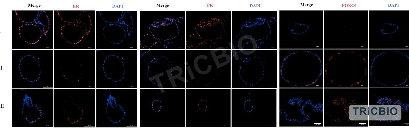 View image
Stage markers: immunofluorescence for ER, PR and FOXO1 adds phenotype context across culture stages for in vitro stage research and candidate-response assessment.
TRiCBIO human endometrial organoid characterization dataStudy planning
Sample and tissue context, the biological objective and the stages or phenotypes of interest guide culture conditions, assays and deliverables.
Sample source, tissue context, cycle or stage information, and available ethics or background documentation.
The tissue-biology objective, induction or intervention conditions, and the stages or phenotypes to compare.
Planned morphology, histology, molecular or functional assays, with preferred images, data and report formats.
Study design
Endometrial organoids support studies of tissue source, epithelial identity and stage-associated change. Each study confirms sample context, culture conditions and the tissue question to be answered.
Assess tissue source, cycle or hormonal context, collection and ethics documentation, then define whether the focus is proliferative, secretory, receptive-associated or treatment-related change.
Use serial bright-field imaging, H&E and E-cadherin/Ki67 profiling to establish model identity and the baseline for stage-focused work.
Define culture points per program and combine ER, PR, FOXO1 and other required assays to assess morphology and molecular change.
Relate baseline model features and stage-dependent biology to treatment-associated changes, then define priorities for functional or translational follow-up.
Research applications
Assays & QC
Baseline characterization combines establishment and passaging morphology, H&E, E-cadherin and Ki67, with ER, PR, FOXO1 or other study-relevant markers used to track stage-dependent change.
Study & delivery
Custom studies define the sample, induction conditions, differentiation stages and assays. Deliverables can include images, results, analysis and a study report.
Related solutions
PROJECT DISCUSSION
Sample and tissue context, study goal, induction or intervention conditions, and stage-specific endpoints determine the model configuration, assay panel and deliverables for the human endometrial organoid.
Track morphology and marker expression from primary establishment and passaging through proliferative, secretory and receptive stages, with epithelial-identity profiling throughout.
Human endometrial tissue, with sample context, cycle stage and applicable ethics requirements incorporated into the study design
3D culture · Stage-directed differentiation
TRiCBIO human endometrial organoid establishment, passage and stage-characterization data
Model characterization
The model dossier covers establishment, passaging, H&E, epithelial identity and stage-related markers, supporting tissue-biology, stage-differentiation and candidate-response studies.
View imageEstablishment and passage: the displayed time points document organoid formation after primary processing and continued growth after passage.
TRiCBIO human endometrial organoid characterization dataView imageBaseline identity: H&E, DAPI, E-cadherin and Ki67 images compare source tissue and organoids for epithelial organization and proliferative state.
TRiCBIO human endometrial organoid characterization dataView imageStage-dependent morphology: serial imaging within one culture workflow helps define time points for downstream assays.
TRiCBIO human endometrial organoid characterization dataView imageStage markers: immunofluorescence for ER, PR and FOXO1 adds phenotype context across culture stages for in vitro stage research and candidate-response assessment.
TRiCBIO human endometrial organoid characterization dataStudy planning
Sample and tissue context, the biological objective and the stages or phenotypes of interest guide culture conditions, assays and deliverables.
Sample source, tissue context, cycle or stage information, and available ethics or background documentation.
The tissue-biology objective, induction or intervention conditions, and the stages or phenotypes to compare.
Planned morphology, histology, molecular or functional assays, with preferred images, data and report formats.
Study design
Endometrial organoids support studies of tissue source, epithelial identity and stage-associated change. Each study confirms sample context, culture conditions and the tissue question to be answered.
Assess tissue source, cycle or hormonal context, collection and ethics documentation, then define whether the focus is proliferative, secretory, receptive-associated or treatment-related change.
Use serial bright-field imaging, H&E and E-cadherin/Ki67 profiling to establish model identity and the baseline for stage-focused work.
Define culture points per program and combine ER, PR, FOXO1 and other required assays to assess morphology and molecular change.
Relate baseline model features and stage-dependent biology to treatment-associated changes, then define priorities for functional or translational follow-up.
Research applications
Assays & QC
Baseline characterization combines establishment and passaging morphology, H&E, E-cadherin and Ki67, with ER, PR, FOXO1 or other study-relevant markers used to track stage-dependent change.
Study & delivery
Custom studies define the sample, induction conditions, differentiation stages and assays. Deliverables can include images, results, analysis and a study report.
Related solutions
PROJECT DISCUSSION
Sample and tissue context, study goal, induction or intervention conditions, and stage-specific endpoints determine the model configuration, assay panel and deliverables for the human endometrial organoid.